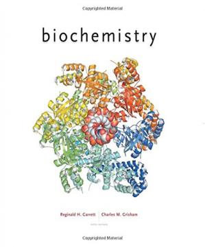
BioChemistry 6th Edition by Reginald Garrett, Charles Grisham 1305577205 9781305577206
$50.00 Original price was: $50.00.$25.00Current price is: $25.00.
BioChemistry 6th Edition by Reginald Garrett, Charles Grisham – Ebook PDF Instant Download/Delivery: 1305577205, 9781305577206
Full download BioChemistry 6th Edition after payment

Product details:
ISBN 10: 1305577205
ISBN 13: 9781305577206
Author: Reginald H Garrett, Charles M Grisham
Continuing Garrett and Grisham’s innovative conceptual and organizing “Essential Questions” framework, BIOCHEMISTRY guides students through course concepts in a way that reveals the beauty and usefulness of biochemistry in the everyday world. Offering a balanced and streamlined presentation, this edition has been updated throughout with new material and revised presentations.
BioChemistry 6th Table of contents:
Part I. Molecular Components of Cells
Chapter 1. The Facts of Life: Chemistry Is the Logic of Biological Phenomena
1.1. What Are the Distinctive Properties of Living Systems?
1.2. What Kinds of Molecules Are Biomolecules?
1.2a. Biomolecules Are Carbon Compounds
1.3. What Is the Structural Organization of Complex Biomolecules?
1.3a. Metabolites Are Used to Form the Building Blocks of Macromolecules
1.3b. Organelles Represent a Higher Order in Biomolecular Organization
1.3c. Membranes Are Supramolecular Assemblies that Define the Boundaries of Cells
1.3d. The Unit of Life Is the Cell
1.4. How Do the Properties of Biomolecules Reflect Their Fitness to the Living Condition?
1.4a. Biological Macromolecules and Their Building Blocks Have a “Sense” or Directionality
1.4b. Biological Macromolecules Are Informational
1.4c. Biomolecules Have Characteristic Three-Dimensional Architecture
1.4d. Weak Forces Maintain Biological Structure and Determine Biomolecular Interactions
1.4e. Van der Waals Attractive Forces Play an Important Role in Biomolecular Interactions
1.4f. Hydrogen Bonds Are Important in Biomolecular Interactions
1.4g. The Defining Concept of Biochemistry Is “Molecular Recognition through Structural Complementarity”
1.4h. Biomolecular Recognition Is Mediated by Weak Chemical Forces
1.4i. Weak Forces Restrict Organisms to a Narrow Range of Environmental Conditions
1.4j. Enzymes Catalyze Metabolic Reactions
1.4k. The Time Scale of Life
1.5. What Are the Organization and Structure of Cells?
1.5a. The Eukaryotic Cell Likely Emerged from an Archaeal Lineage
1.5b. How Many Genes Does a Cell Need?
1.5c. Archaea and Bacteria Have a Relatively Simple Structural Organization
1.5d. The Structural Organization of Eukaryotic Cells Is More Complex than That of Prokaryotic Cells
1.6. What Are Viruses?
Summary
Foundational Biochemistry
Problems
Further Reading
Chapter 2. Water: The Medium of Life
2.1. What Are the Properties of Water?
2.1a. Water Has Unusual Properties
2.1b. Hydrogen Bonding in Water Is Key to Its Properties
2.1c. The Structure of Ice Is Based on H -Bond Formation
2.1d. Molecular Interactions in Liquid Water Are Based on H Bonds
2.1e. The Solvent Properties of Water Derive from Its Polar Nature
2.1f. Water Can Ionize to Form H + and OH –
2.2. What Is pH?
2.2a. Strong Electrolytes Dissociate Completely in Water
2.2b. Weak Electrolytes Are Substances that Dissociate Only Slightly in Water
2.2c. The Henderson–Hasselbalch Equation Describes the Dissociation of a Weak Acid in the Presence of Its Conjugate Base
2.2d. Titration Curves Illustrate the Progressive Dissociation of a Weak Acid
2.2e. Phosphoric Acid Has Three Dissociable H +
2.3. What Are Buffers, and What Do They Do?
2.3a. The Phosphate Buffer System Is a Major Intracellular Buffering System
2.3b. The Imidazole Group of Histidine Also Serves as an Intracellular Buffering System
2.3c. “Good” Buffers Are Buffers Useful within Physiological pH Ranges
2.4. What Properties of Water Give It a Unique Role in the Environment?
Summary
Foundational Biochemistry
Problems
Further Reading
Chapter 3. Thermodynamics of Biological Systems
3.1. What Are the Basic Concepts of Thermodynamics?
3.1a. Three Quantities Describe the Energetics of Biochemical Reactions
3.1b. All Reactions and Processes Follow the Laws of Thermodynamics
3.1c. Free Energy Provides a Simple Criterion for Equilibrium
3.2. What Is the Effect of Concentration on Net Free Energy Changes?
3.3. What Is the Effect of pH on Standard-State Free Energies?
3.4. What Can Thermodynamic Parameters Tell Us about Biochemical Events?
3.5. What Are the Characteristics of High-Energy Biomolecules?
3.5a. ATP Is an Intermediate Energy-Shuttle Molecule
3.5b. Group Transfer Potentials Quantify the Reactivity of Functional Groups
3.5c. The Hydrolysis of Phosphoric Acid Anhydrides Is Highly Favorable
3.5d. The Hydrolysis Δ G ° ′ of ATP and ADP Is Greater than That of AMP
3.5e. Acetyl Phosphate and 1,3-Bisphosphoglycerate Are Phosphoric-Carboxylic Anhydrides
3.5f. Enol Phosphates Are Potent Phosphorylating Agents
3.6. What Are the Complex Equilibria Involved in ATP Hydrolysis?
3.6a. The Δ G ° ′ of Hydrolysis for ATP Is pH-Dependent
3.6b. Metal Ions Affect the Free Energy of Hydrolysis of ATP
3.6c. Concentration Affects the Free Energy of Hydrolysis of ATP
3.7. Why Are Coupled Processes Important to Living Things?
3.8. What Is the Daily Human Requirement for ATP?
3.9. What Are Reduction Potentials, and How Are They Used to Account for Free Energy Changes in Redox Reactions?
3.9a. Standard Reduction Potentials Are Measured in Reaction Half-Cells
3.9b. ℰ o ′ Values Can Be Used to Predict the Direction of Redox Reactions
3.9c. ℰ o ′ Values Can Be Used to Analyze Energy Changes in Redox Reactions
3.9d. The Reduction Potential Depends on Concentration
Summary
Foundational Biochemistry
Problems
Further Reading
Chapter 4. Amino Acids and the Peptide Bond
4.1. What Are the Structures and Properties of Amino Acids?
4.1a. Typical Amino Acids Contain a Central Tetrahedral Carbon Atom
4.1b. Amino Acids Can Join via Peptide Bonds
4.1c. There Are 20 Common Amino Acids
4.1d. Are There Other Ways to Classify Amino Acids?
4.1e. Amino Acids 21 and 22 —and More?
4.1f. Several Amino Acids Occur Only Rarely in Proteins
4.2. What Are the Acid–Base Properties of Amino Acids?
4.2a. Amino Acids Are Weak Polyprotic Acids
4.2b. Side Chains of Amino Acids Undergo Characteristic Ionizations
4.3. What Reactions Do Amino Acids Undergo?
4.4. What Are the Optical and Stereochemical Properties of Amino Acids?
4.4a. Amino Acids Are Chiral Molecules
4.4b. Chiral Molecules Are Described by the D , L and ( R , S ) Naming Conventions
4.5. What Are the Spectroscopic Properties of Amino Acids?
4.5a. Phenylalanine, Tyrosine, and Tryptophan Absorb Ultraviolet Light
4.5b. Amino Acids Can Be Characterized by Nuclear Magnetic Resonance
4.6. How Are Amino Acid Mixtures Separated and Analyzed?
4.6a. Amino Acids Can Be Separated by Chromatography
4.7. What Is the Fundamental Structural Pattern in Proteins?
4.7a. The Peptide Bond Has Partial Double-Bond Character
4.7b. The Polypeptide Backbone Is Relatively Polar
4.7c. Peptides Can Be Classified According to How Many Amino Acids They Contain
4.7d. Proteins Are Composed of One or More Polypeptide Chains
Summary
Foundational Biochemistry
Problems
Further Reading
Chapter 5. Proteins: Their Primary Structure and Biological Functions
5.1. What Architectural Arrangements Characterize Protein Structure?
5.1a. Proteins Fall into Three Basic Classes According to Shape and Solubility
5.1b. Protein Structure Is Described in Terms of Four Levels of Organization
5.1c. Noncovalent Forces Drive Formation of the Higher Orders of Protein Structure
5.1d. A Protein’s Conformation Can Be Described as Its Overall Three-Dimensional Structure
5.2. How Are Proteins Isolated and Purified from Cells?
5.2a. A Number of Protein Separation Methods Exploit Differences in Size and Charge
5.2b. A Typical Protein Purification Scheme Uses a Series of Separation Methods
5.3. How Is the Amino Acid Analysis of Proteins Performed?
5.3a. Acid Hydrolysis Liberates the Amino Acids of a Protein
5.3b. Chromatographic Methods Are Used to Separate the Amino Acids
5.3c. The Amino Acid Compositions of Different Proteins Are Different
5.4. How Is the Primary Structure of a Protein Determined?
5.4a. The Sequence of Amino Acids in a Protein Is Distinctive
5.4b. Sanger Was the First to Determine the Sequence of a Protein
5.4c. Both Chemical and Enzymatic Methodologies Are Used in Protein Sequencing
5.4d. Step 1. Separation of Polypeptide Chains
5.4e. Step 2. Cleavage of Disulfide Bridges
5.4f. Step 3. N- and C-Terminal Analysis
5.4g. Steps 4 and 5. Fragmentation of the Polypeptide Chain
5.4h. Step 6. Reconstruction of the Overall Amino Acid Sequence
5.4i. The Amino Acid Sequence of a Protein Can Be Determined by Mass Spectrometry
5.4j. Sequence Databases Contain the Amino Acid Sequences of Millions of Different Proteins
5.5. What Is the Nature of Amino Acid Sequences?
5.5a. Homologous Proteins from Different Organisms Have Homologous Amino Acid Sequences
5.5b. Computer Programs Can Align Sequences and Discover Homology between Proteins
5.5c. Related Proteins Share a Common Evolutionary Origin
5.5d. Apparently Different Proteins May Share a Common Ancestry
5.5e. A Mutant Protein Is a Protein with a Slightly Different Amino Acid Sequence
5.6. Can Polypeptides Be Synthesized in the Laboratory?
5.6a. Solid-Phase Methods Are Very Useful in Peptide Synthesis
5.7. Do Proteins Have Chemical Groups Other than Amino Acids?
5.8. What Are the Many Biological Functions of Proteins?
All Proteins Function through Specific Recognition and Binding of Some Target Molecule
5.9. What Is the Proteome and What Does It Tell Us?
5.9a. The Proteome Is Dynamic
5.9b. Determining the Proteome of a Cell
Summary
Foundational Biochemistry
Problems
Further Reading
Chapter 6. Proteins: Secondary, Tertiary, and Quaternary Structure
6.1. What Noncovalent Interactions Stabilize the Higher Levels of Protein Structure?
6.1a. Hydrogen Bonds Are Formed whenever Possible
6.1b. Hydrophobic Interactions Drive Protein Folding
6.1c. Ionic Interactions Usually Occur on the Protein Surface
6.1d. Van der Waals Interactions Are Ubiquitous
6.2. What Role Does the Amino Acid Sequence Play in Protein Structure?
6.3. What Are the Elements of Secondary Structure in Proteins, and How Are They Formed?
6.3a. All Protein Structure Is Based on the Amide Plane
6.3b. The Alpha-Helix Is a Key Secondary Structure
6.3c. The β -Pleated Sheet Is a Core Structure in Proteins
6.3d. Helix–Sheet Composites in Spider Silk
6.3e. β -Turns Allow the Protein Strand to Change Direction
6.4. How Do Polypeptides Fold into Three-Dimensional Protein Structures?
6.4a. Fibrous Proteins Usually Play a Structural Role
6.4b. Globular Proteins Mediate Cellular Function
6.4c. Helices and Sheets Make up the Core of Most Globular Proteins
6.4d. Waters on the Protein Surface Stabilize the Structure
6.4e. Packing Considerations
6.4f. Protein Domains Are Nature’s Modular Strategy for Protein Design
6.4g. Classification Schemes for the Protein Universe Are Based on Domains
6.4h. Denaturation Leads to Loss of Protein Structure and Function
6.4i. Anfinsen’s Classic Experiment Proved that Sequence Determines Structure
6.4j. Is There a Single Mechanism for Protein Folding?
6.4k. What Is the Thermodynamic Driving Force for Folding of Globular Proteins?
6.4l. Marginal Stability of the Tertiary Structure Makes Proteins Flexible
6.4m. Motion in Globular Proteins
6.4n. The Folding Tendencies and Patterns of Globular Proteins
6.4o. Most Globular Proteins Belong to One of Four Structural Classes
6.4p. Molecular Chaperones Are Proteins that Help other Proteins to Fold
6.4q. Some Proteins Are Intrinsically Unstructured
6.5. How Do Protein Subunits Interact at the Quaternary Level of Protein Structure?
6.5a. There Is Symmetry in Quaternary Structures
6.5b. Quaternary Association Is Driven by Weak Forces
6.5c. Open Quaternary Structures Can Polymerize
6.5d. There Are Structural and Functional Advantages to Quaternary Association
Summary
Foundational Biochemistry
Problems
Further Reading
Chapter 7. Carbohydrates and the Glycoconjugates of Cell Surfaces
7.1. How Are Carbohydrates Named?
7.2. What Are the Structure and Chemistry of Monosaccharides?
7.2a. Monosaccharides Are Classified as Aldoses and Ketoses
7.2b. Stereochemistry Is a Prominent Feature of Monosaccharides
7.2c. Monosaccharides Exist in Cyclic and Anomeric Forms
7.2d. Haworth Projections Are a Convenient Device for Drawing Sugars
7.2e. Monosaccharides Can Be Converted to Several Derivative Forms
7.3. What Are the Structure and Chemistry of Oligosaccharides?
7.3a. Disaccharides Are the Simplest Oligosaccharides
7.3b. A Variety of Higher Oligosaccharides Occur in Nature
7.4. What Are the Structure and Chemistry of Polysaccharides?
7.4a. Nomenclature for Polysaccharides Is Based on Their Composition and Structure
7.4b. Polysaccharides Serve Energy Storage, Structure, and Protection Functions
7.4c. Polysaccharides Provide Stores of Energy
7.4d. Polysaccharides Provide Physical Structure and Strength to Organisms
7.4e. Polysaccharides Provide Strength and Rigidity to Bacterial Cell Walls
7.4f. Peptidoglycan Is the Polysaccharide of Bacterial Cell Walls
7.4g. Animals Display a Variety of Cell Surface Polysaccharides
7.5. What Are Glycoproteins, and How Do They Function in Cells?
7.5a. Carbohydrates on Proteins Can Be O-Linked or N-Linked
7.5b. O-GlcNAc Signaling Is Altered in Diabetes and Cancer
7.5c. O-Linked Saccharides Form Rigid Extended Extracellular Structures
7.5d. Polar Fish Depend on Antifreeze Glycoproteins
7.5e. N-Linked Oligosaccharides Can Affect the Physical Properties and Functions of a Protein
7.5f. Sialic Acid Terminates the Oligosaccharides of Glycoproteins and Glycolipids
7.5g. Sialic Acid Cleavage Can Serve as a Timing Device for Protein Degradation
7.6. How Do Proteoglycans Modulate Processes in Cells and Organisms?
7.6a. Functions of Proteoglycans Involve Binding to Other Proteins
7.6b. Proteoglycans May Modulate Cell Growth Processes
7.6c. Proteoglycans Make Cartilage Flexible and Resilient
7.7. Do Carbohydrates Provide a Structural Code?
7.7a. Lectins Translate the Sugar Code
7.7b. Selectins, Rolling Leukocytes, and the Inflammatory Response
7.7c. Galectins—Mediators of Inflammation, Immunity, and Cancer
7.7d. C-Reactive Protein—A Lectin that Limits Inflammation Damage
Summary
Foundational Biochemistry
Problems
Further Reading
Chapter 8. Lipids
8.1. What Are the Structures and Chemistry of Fatty Acids?
8.2. What Are the Structures and Chemistry of Triacylglycerols?
8.3. What Are the Structures and Chemistry of Glycerophospholipids?
8.3a. Glycerophospholipids Are the Most Common Phospholipids
8.3b. Ether Glycerophospholipids Include PAF and Plasmalogens
8.4. What Are Sphingolipids, and How Are They Important for Higher Animals?
8.5. What Are Waxes, and How Are They Used?
8.6. What Are Terpenes, and What Is Their Relevance to Biological Systems?
The Membranes of Archaea Are Rich in Isoprene-Based Lipids
8.7. What Are Steroids, and What Are Their Cellular Functions?
8.7a. Cholesterol
8.7b. Steroid Hormones Are Derived from Cholesterol
8.8. How Do Lipids and Their Metabolites Act as Biological Signals?
8.9. What Can Lipidomics Tell Us about Cell, Tissue, and Organ Physiology?
Summary
Foundational Biochemistry
Problems
Further Reading
Chapter 9. Membranes and Membrane Transport
9.1. What Are the Chemical and Physical Properties of Membranes?
9.1a. The Composition of Membranes Suits Their Functions
9.1b. Lipids Form Ordered Structures Spontaneously in Water
9.1c. The Fluid Mosaic Model Describes Membrane Dynamics
9.2. What Are the Structure and Chemistry of Membrane Proteins?
9.2a. Peripheral Membrane Proteins Associate Loosely with the Membrane
9.2b. Integral Membrane Proteins Are Firmly Anchored in the Membrane
9.2c. Lipid-Anchored Membrane Proteins Are Switching Devices
9.3. How Are Biological Membranes Organized?
9.3a. Membranes Are Asymmetric and Heterogeneous Structures
9.4. What Are the Dynamic Processes That Modulate Membrane Function?
9.4a. Lipids and Proteins Undergo a Variety of Movements in Membranes
9.4b. Membrane Lipids Can Be Ordered to Different Extents
9.5. How Does Transport Occur across Biological Membranes?
9.6. What Is Passive Diffusion?
9.6a. Charged Species May Cross Membranes by Passive Diffusion
9.7. How Does Facilitated Diffusion Occur?
9.7a. Membrane Channel Proteins Facilitate Diffusion
9.7b. The B. cereus Na K Channel Uses a Variation on the K + Selectivity Filter
9.7c. CorA Is a Pentameric Mg 2 + Channel
9.7d. Chloride, Water, Glycerol, and Ammonia Flow through Single-Subunit Pores
9.8. How Does Energy Input Drive Active Transport Processes?
9.8a. All Active Transport Systems Are Energy-Coupling Devices
9.8b. Many Active Transport Processes Are Driven by ATP
9.8c. ABC Transporters Use ATP to Drive Import and Export Functions and Provide Multidrug Resistance
9.9. How Are Certain Transport Processes Driven by Light Energy?
9.9a. Bacteriorhodopsin Uses Light Energy to Drive Proton Transport
9.10. How Is Secondary Active Transport Driven by Ion Gradients?
9.10a. Na + and H + Drive Secondary Active Transport
9.10b. AcrB Is a Secondary Active Transport System
Summary
Foundational Biochemistry
Problems
Further Reading
Chapter 10. Nucleotides and Nucleic Acids
10.1. What Are the Structure and Chemistry of Nitrogenous Bases?
10.1a. Three Pyrimidines and Two Purines Are Commonly Found in Cells
10.1b. The Properties of Pyrimidines and Purines Can Be Traced to Their Electron-Rich Nature
10.2. What Are Nucleosides?
10.3. What Are the Structure and Chemistry of Nucleotides?
10.3a. Cyclic Nucleotides Are Cyclic Phosphodiesters
10.3b. Nucleoside Diphosphates and Triphosphates Are Nucleotides with Two or Three Phosphate Groups
10.3c. NDPs and NTPs Are Polyprotic Acids
10.3d. Nucleoside 5 ′ -Triphosphates Are Carriers of Chemical Energy
10.4. What Are Nucleic Acids?
10.4a. The Base Sequence of a Nucleic Acid Is Its Defining Characteristic
10.5. What Are the Different Classes of Nucleic Acids?
10.5a. The Fundamental Structure of DNA Is a Double Helix
10.5b. Various Forms of RNA Serve Different Roles in Cells
10.5c. The Chemical Differences between DNA and RNA Have Biological Significance
10.6. Are Nucleic Acids Susceptible to Hydrolysis?
10.6a. RNA Is Susceptible to Hydrolysis by Base, but DNA Is Not
10.6b. The Enzymes that Hydrolyze Nucleic Acids Are Phosphodiesterases
10.6c. Nucleases Differ in Their Specificity for Different Forms of Nucleic Acid
10.6d. Restriction Enzymes Are Nucleases that Cleave Double-Stranded DNA Molecules
10.6e. Type II Restriction Endonucleases Are Useful for Manipulating DNA in the Lab
10.6f. Restriction Endonucleases Can Be Used to Map the Structure of a DNA Fragment
Summary
Foundational Biochemistry
Problems
Further Reading
Chapter 11. Structure of Nucleic Acids
11.1. How Do Scientists Determine the Primary Structure of Nucleic Acids?
11.1a. The Nucleotide Sequence of DNA Can Be Determined from the Electrophoretic Migration of a Defined Set of Polynucleotide Fragments
11.1b. Sanger’s Chain Termination or Dideoxy Method Uses DNA Replication to Generate a Defined Set of Polynucleotide Fragments
11.1c. Next-Generation Sequencing
11.1d. High-Throughput DNA Sequencing by the Light of Fireflies
11.1e. Illumina Next-Gen Sequencing
11.1f. Emerging Technologies to Sequence DNA Are Based on Single-Molecule Sequencing Strategies
11.2. What Sorts of Secondary Structures Can Double-Stranded DNA Molecules Adopt?
11.2a. Conformational Variation in Polynucleotide Strands
11.2b. DNA Usually Occurs in the Form of Double-Stranded Molecules
11.2c. Watson–Crick Base Pairs Have Virtually Identical Dimensions
11.2d. The DNA Double Helix Is a Stable Structure
11.2e. Double Helical Structures Can Adopt a Number of Stable Conformations
11.2f. A-Form DNA Is an Alternative Form of Right-Handed DNA
11.2g. Z-DNA Is a Conformational Variation in the Form of a Left-Handed Double Helix
11.2h. The Double Helix Is a Very Dynamic Structure
11.2i. Alternative Hydrogen-Bonding Interactions Give Rise to Novel DNA Structures: Cruciforms, Triplexes, and Quadruplexes
11.3. Can the Secondary Structure of DNA Be Denatured and Renatured?
11.3a. Thermal Denaturation of DNA Can Be Observed by Changes in UV Absorbance
11.3b. pH Extremes or Strong H -Bonding Solutes also Denature DNA Duplexes
11.3c. Single-Stranded DNA Can Renature to Form DNA Duplexes
11.3d. The Rate of DNA Renaturation Is an Index of DNA Sequence Complexity
11.3e. Nucleic Acid Hybridization: Different DNA Strands of Similar Sequence Can Form Hybrid Duplexes
11.4. Can DNA Adopt Structures of Higher Complexity?
11.4a. Supercoils Are One Kind of Structural Complexity in DNA
11.5. What Is the Structure of Eukaryotic Chromosomes?
11.5a. Nucleosomes Are the Fundamental Structural Unit in Chromatin
11.5b. Higher-Order Structural Organization of Chromatin Gives Rise to Chromosomes
11.5c. SMC Proteins Establish Chromosome Organization and Mediate Chromosome Dynamics
11.6. Can Nucleic Acids Be Synthesized Chemically?
11.6a. Phosphoramidite Chemistry Is Used to Form Oligonucleotides from Nucleotides
11.6b. Genes Can Be Synthesized Chemically
11.7. What Are the Secondary and Tertiary Structures of RNA?
11.7a. Transfer RNA Adopts Higher-Order Structure through Intrastrand Base Pairing
11.7b. Messenger RNA Adopts Higher-Order Structure through Intrastrand Base Pairing
11.7c. Ribosomal RNA Also Adopts Higher-Order Structure through Intrastrand Base Pairing
11.7d. Aptamers Are Oligonucleotides Specifically Selected for Their Ligand-Binding Ability
Summary
Foundational Biochemistry
Problems
Further Reading
Chapter 12. Recombinant DNA, Cloning, Chimeric Genes, and Synthetic Biology
12.1. What Does It Mean “To Clone”?
12.1a. Plasmids Are Very Useful in Cloning Genes
12.1b. Shuttle Vectors Are Plasmids that Can Propagate in Two Different Organisms
12.1c. Artificial Chromosomes Can Be Created from Recombinant DNA
12.2. What Is a DNA Library?
12.2a. Genomic Libraries Are Prepared from the Total DNA in an Organism
12.2b. Libraries Can Be Screened for the Presence of Specific Genes
12.2c. Probes for Screening Libraries Can Be Prepared in a Variety of Ways
12.2d. PCR Is Used to Clone and Amplify Specific Genes
12.2e. cDNA Libraries Are DNA Libraries Prepared from mRNA
12.2f. DNA Microarrays (Gene Chips) Are Arrays of Different Oligonucleotides Immobilized on a Chip
12.3. Can the Cloned Genes in Libraries Be Expressed?
12.3a. Expression Vectors Are Engineered so that the RNA or Protein Products of Cloned Genes Can Be Expressed
12.3b. Reporter Gene Constructs Are Chimeric DNA Molecules Composed of Gene Regulatory Sequences Positioned next to an Easily Expressible Gene Product
12.3c. Specific Protein–Protein Interactions Can Be Identified Using the Yeast Two-Hybrid System
12.3d. In Vitro Mutagenesis
12.4. How Is RNA Interference Used to Reveal the Function of Genes?
12.4a. RNAi Using Synthetic shRNAs
12.5. How Does High-Throughput Technology Allow Global Study of Millions of Genes or Molecules at Once?
12.5a. High-Throughput Screening
12.5b. DNA Laser Printing
12.5c. High-Throughput RNAi Screening of Mammalian Genomes
12.5d. High-Throughput Protein Screening
12.6. Is It Possible to Make Directed Changes in the Heredity of an Organism?
12.6a. Human Gene Therapy Can Repair Genetic Deficiencies
12.6b. Viruses as Vectors in Human Gene Therapy
12.7. What Is the New Field of Synthetic Biology?
12.7a. DNA as Code
12.7b. iGEM and BioBricks (Registry of Standard Biological Parts)
12.7c. Metabolic Engineering
12.7d. Genome Engineering
12.7e. Genome Editing with CRISPR/Cas9
12.7f. Synthetic Genomes
Summary
Foundational Biochemistry
Problems
Further Reading
Part II. Protein Dynamics
Chapter 13. Enzymes—Kinetics and Specificity
13.1. What Characteristic Features Define Enzymes?
13.1a. Catalytic Power Is Defined as the Ratio of the Enzyme-Catalyzed Rate of a Reaction to the Uncatalyzed Rate
13.1b. Specificity Is the Term Used to Define the Selectivity of Enzymes for Their Substrates
13.1c. Regulation of Enzyme Activity Ensures That the Rate of Metabolic Reactions Is Appropriate to Cellular Requirements
13.1d. Enzyme Nomenclature Provides a Systematic Way of Naming Metabolic Reactions
13.1e. Coenzymes and Cofactors Are Nonprotein Components Essential to Enzyme Activity
13.2. Can the Rate of an Enzyme-Catalyzed Reaction Be Defined in a Mathematical Way?
13.2a. Chemical Kinetics Provides a Foundation for Exploring Enzyme Kinetics
13.2b. Bimolecular Reactions Are Reactions Involving Two Reactant Molecules
13.2c. Catalysts Lower the Free Energy of Activation for a Reaction
13.2d. Decreasing Δ G ‡ Increases Reaction Rate
13.3. What Equations Define the Kinetics of Enzyme-Catalyzed Reactions?
13.3a. The Substrate Binds at the Active Site of an Enzyme
13.3b. The Michaelis–Menten Equation Is the Fundamental Equation of Enzyme Kinetics
13.3c. Assume That [ES] Remains Constant during an Enzymatic Reaction
13.3d. Assume That Velocity Measurements Are Made Immediately after Adding S
13.3e. The Michaelis Constant, K m , Is Defined as ( k – 1 + k 2 ) / k 1
13.3f. When [ S ] = K m , v = V max / 2
13.3g. Plots of v versus [S] Illustrate the Relationships between V max , K m , and Reaction Order
13.3h. Turnover Number Defines the Activity of One Enzyme Molecule
13.3i. The Ratio, k cat / K m , Defines the Catalytic Efficiency of an Enzyme
13.3j. Linear Plots Can Be Derived from the Michaelis–Menten Equation
13.3k. Nonlinear Lineweaver–Burk or Hanes–Woolf Plots Are a Property of Regulatory Enzymes
13.3l. Enzymatic Activity Is Strongly Influenced by pH
13.3m. The Response of Enzymatic Activity to Temperature Is Complex
13.4. What Can Be Learned from the Inhibition of Enzyme Activity?
13.4a. Enzymes May Be Inhibited Reversibly or Irreversibly
13.4b. Reversible Inhibitors May Bind at the Active Site or at Some Other Site
13.4c. Enzymes Also Can Be Inhibited in an Irreversible Manner
13.5. What Is the Kinetic Behavior of Enzymes Catalyzing Bimolecular Reactions?
13.5a. The Conversion of AEB to PEQ Is the Rate-Limiting Step in Random, Single-Displacement Reactions
13.5b. In an Ordered, Single-Displacement Reaction, the Leading Substrate Must Bind First
13.5c. Double-Displacement (Ping-Pong) Reactions Proceed via Formation of a Covalently Modified Enzyme Intermediate
13.5d. Exchange Reactions Are One Way to Diagnose Bisubstrate Mechanisms
13.5e. Multisubstrate Reactions Can Also Occur in Cells
13.6. How Can Enzymes Be so Specific?
13.6a. The “Lock and Key” Hypothesis Was the First Explanation for Specificity
13.6b. The “Induced Fit” Hypothesis Provides a More Accurate Description of Specificity
13.6c. “Induced Fit” Favors Formation of the Transition State
13.6d. Specificity and Reactivity
13.7. Are All Enzymes Proteins?
13.7a. RNA Molecules That Are Catalytic Have Been Termed “Ribozymes”
13.7b. Antibody Molecules Can Have Catalytic Activity
13.8. Is It Possible to Design an Enzyme to Catalyze Any Desired Reaction?
Summary
Foundational Biochemistry
Problems
Further Reading
Chapter 14. Mechanisms of Enzyme Action
14.1. What Are the Magnitudes of Enzyme-Induced Rate Accelerations?
14.2. What Role Does Transition-State Stabilization Play in Enzyme Catalysis?
14.3. How Does Destabilization of ES Affect Enzyme Catalysis?
14.4. How Tightly Do Transition-State Analogs Bind to the Active Site?
14.5. What Are the Mechanisms of Catalysis?
14.5a. Enzymes Facilitate Formation of Near-Attack Conformations
14.5b. Protein Motions Are Essential to Enzyme Catalysis
14.5c. Covalent Catalysis
14.5d. General Acid–Base Catalysis
14.5e. Low-Barrier Hydrogen Bonds
14.5f. Quantum Mechanical Tunneling in Electron and Proton Transfers
14.5g. Metal Ion Catalysis
14.6. What Can Be Learned from Typical Enzyme Mechanisms?
14.6a. Serine Proteases
14.6b. The Digestive Serine Proteases
14.6c. The Chymotrypsin Mechanism in Detail: Kinetics
14.6d. The Serine Protease Mechanism in Detail: Events at the Active Site
14.6e. The Aspartic Proteases
14.6f. The Mechanism of Action of Aspartic Proteases
14.6g. The AIDS Virus HIV-1 Protease Is an Aspartic Protease
14.6h. Chorismate Mutase: A Model for Understanding Catalytic Power and Efficiency
Summary
Foundational Biochemistry
Problems
Further Reading
Chapter 15. Enzyme Regulation
15.1. What Factors Influence Enzymatic Activity?
15.1a. The Availability of Substrates and Cofactors Usually Determines How Fast the Reaction Goes
15.1b. As Product Accumulates, the Apparent Rate of the Enzymatic Reaction Will Decrease
15.1c. Genetic Regulation of Enzyme Synthesis and Decay Determines the Amount of Enzyme Present at Any Moment
15.1d. Enzyme Activity Can Be Regulated Allosterically
15.1e. Enzyme Activity Can Be Regulated through Covalent Modification
15.1f. Regulation of Enzyme Activity Also Can Be Accomplished in Other Ways
15.1g. Zymogens Are Inactive Precursors of Enzymes
15.1h. Isozymes Are Enzymes with Slightly Different Subunits
15.2. What Are the General Features of Allosteric Regulation?
15.2a. Regulatory Enzymes Have Certain Exceptional Properties
15.3. Can Allosteric Regulation Be Explained by Conformational Changes in Proteins?
15.3a. The Symmetry Model for Allosteric Regulation Is Based on Two Conformational States for a Protein
15.3b. The Sequential Model for Allosteric Regulation Is Based on Ligand-Induced Conformational Changes
15.3c. Changes in the Oligomeric State of a Protein Can Also Give Allosteric Behavior
15.4. What Kinds of Covalent Modification Regulate the Activity of Enzymes?
15.4a. Covalent Modification through Reversible Phosphorylation
15.4b. Protein Kinases: Target Recognition and Intrasteric Control
15.4c. Phosphorylation Is Not the Only Form of Covalent Modification That Regulates Protein Function
15.4d. Acetylation Is a Prominent Modification for the Regulation of Metabolic Enzymes
15.5. Is the Activity of Some Enzymes Controlled by both Allosteric Regulation and Covalent Modification?
15.5a. The Glycogen Phosphorylase Reaction Converts Glycogen into Readily Usable Fuel in the Form of Glucose-1-Phosphate
15.5b. Glycogen Phosphorylase Is a Homodimer
15.5c. Glycogen Phosphorylase Activity Is Regulated Allosterically
15.5d. Covalent Modification of Glycogen Phosphorylase Trumps Allosteric Regulation
15.5e. Enzyme Cascades Regulate Glycogen Phosphorylase Covalent Modification
Special Focus. Is There an Example in Nature That Exemplifies the Relationship between Quaternary Structure and the Emergence of Allosteric Properties? Hemoglobin and Myoglobin—Paradigms of Protein Structure and Function
The Comparative Biochemistry of Myoglobin and Hemoglobin Reveals Insights into Allostery
Myoglobin Is an Oxygen-Storage Protein
O 2 Binds to the Mb Heme Group
O 2 Binding Alters Mb Conformation
Cooperative Binding of Oxygen by Hemoglobin Has Important Physiological Significance
Hemoglobin Has an α 2 β 2 Tetrameric Structure
Oxygenation Markedly Alters the Quaternary Structure of Hb
Movement of the Heme Iron by less than 0.04 nm Induces the Conformational Change in Hemoglobin
The Oxy and Deoxy Forms of Hemoglobin Represent Two Different Conformational States
The Allosteric Behavior of Hemoglobin Has both Symmetry (MWC) Model and Sequential (KNF) Model Components
H + Promotes the Dissociation of Oxygen from Hemoglobin
CO 2 Also Promotes the Dissociation of O 2 from Hemoglobin
2,3-Bisphosphoglycerate Is an Important Allosteric Effector for Hemoglobin
BPG Binding to Hb Has Important Physiological Significance
Fetal Hemoglobin Has a Higher Affinity for O 2 because It Has a Lower Affinity for BPG
Sickle-Cell Anemia Is Characterized by Abnormal Red Blood Cells
Sickle-Cell Anemia Is a Molecular Disease
Summary
Foundational Biochemistry
Problems
Further Reading
Chapter 16. Molecular Motors
16.1. What Is a Molecular Motor?
16.2. What Is the Molecular Mechanism of Muscle Contraction?
16.2a. Muscle Contraction Is Triggered by Ca 2 + Release from Intracellular Stores
16.2b. The Molecular Structure of Skeletal Muscle Is Based on Actin and Myosin
16.2c. The Mechanism of Muscle Contraction Is Based on Sliding Filaments
16.3. What Are the Molecular Motors That Orchestrate the Mechanochemistry of Microtubules?
16.3a. Filaments of the Cytoskeleton Are Highways That Move Cellular Cargo
16.3b. Three Classes of Motor Proteins Move Intracellular Cargo
16.3c. Dyneins Move Organelles in a Plus-to-Minus Direction; Kinesins, in a Minus-to-Plus Direction—Mostly
16.3d. Cytoskeletal Motors Are Highly Processive
16.3e. ATP Binding and Hydrolysis Drive Hand-over-Hand Movement of Kinesin
16.3f. The Conformation Change That Leads to Movement Is Different in Myosins and Dyneins
16.4. How Do Molecular Motors Unwind DNA?
16.4a. Negative Cooperativity Facilitates Hand-over-Hand Movement
16.4b. Papillomavirus E1 Helicase Moves along DNA on a Spiral Staircase
16.5. How Do Bacterial Flagella Use a Proton Gradient to Drive Rotation?
16.5a. The Flagellar Rotor Is a Complex Structure
16.5b. Gradients of H + and Na + Drive Flagellar Rotors
16.5c. The Flagellar Rotor Self-Assembles in a Spontaneous Process
16.5d. Flagellar Filaments Are Composed of Protofilaments of Flagellin
16.5e. Motor Reversal Involves Conformation Switching of Motor and Filament Proteins
Summary
Foundational Biochemistry
Problems
Further Reading
Part III. Metabolism and Its Regulation
Chapter 17. Metabolism: An Overview
17.1. Is Metabolism Similar in Different Organisms?
17.1a. Living Things Exhibit Metabolic Diversity
17.1b. Oxygen Is Essential to Life for Aerobes
17.1c. The Flow of Energy in the Biosphere and the Carbon and Oxygen Cycles Are Intimately Related
17.2. What Can Be Learned from Metabolic Maps?
17.2a. The Metabolic Map Can Be Viewed as a Set of Dots and Lines
17.2b. Alternative Models Can Provide New Insights into Pathways
17.2c. Multienzyme Systems May Take Different Forms
17.3. How Do Anabolic and Catabolic Processes Form the Core of Metabolic Pathways?
17.3a. Anabolism Is Biosynthesis
17.3b. Anabolism and Catabolism Are Not Mutually Exclusive
17.3c. The Pathways of Catabolism Converge to a Few End Products
17.3d. Anabolic Pathways Diverge, Synthesizing an Astounding Variety of Biomolecules from a Limited Set of Building Blocks
17.3e. Amphibolic Intermediates Play Dual Roles
17.3f. Corresponding Pathways of Catabolism and Anabolism Differ in Important Ways
17.3g. ATP Serves in a Cellular Energy Cycle
17.3h. NAD + Collects Electrons Released in Catabolism
17.3i. NADPH Provides the Reducing Power for Anabolic Processes
17.3j. Coenzymes and Vitamins Provide Unique Chemistry and Essential Nutrients to Pathways
17.4. What Experiments Can Be Used to Elucidate Metabolic Pathways?
17.4a. Mutations Create Specific Metabolic Blocks
17.4b. Isotopic Tracers Can Be Used as Metabolic Probes
17.4c. NMR Spectroscopy Is a Noninvasive Metabolic Probe
17.4d. Metabolic Pathways Are Compartmentalized within Cells
17.5. What Can the Metabolome Tell Us about a Biological System?
Discovery Metabolomics
17.6. What Food Substances Form the Basis of Human Nutrition?
17.6a. Humans Require Protein
17.6b. Carbohydrates Provide Metabolic Energy
17.6c. Lipids Are Essential, but in Moderation
Summary
Foundational Biochemistry
Problems
Further Reading
Chapter 18. Glycolysis
18.1. What Are the Essential Features of Glycolysis?
18.2. Why Are Coupled Reactions Important in Glycolysis?
18.3. What Are the Chemical Principles and Features of the First Phase of Glycolysis?
18.3a. Reaction 1: Glucose Is Phosphorylated by Hexokinase or Glucokinase—The First Priming Reaction
18.3b. Reaction 2: Phosphoglucoisomerase Catalyzes the Isomerization of Glucose-6-Phosphate
18.3c. Reaction 3: ATP Drives a Second Phosphorylation by Phosphofructokinase—The Second Priming Reaction
18.3d. Reaction 4: Cleavage by Fructose Bisphosphate Aldolase Creates Two 3-Carbon Intermediates
18.3e. Reaction 5: Triose Phosphate Isomerase Completes the First Phase of Glycolysis
18.4. What Are the Chemical Principles and Features of the Second Phase of Glycolysis?
18.4a. Reaction 6: Glyceraldehyde-3-Phosphate Dehydrogenase Creates a High-Energy Intermediate
18.4b. Reaction 7: Phosphoglycerate Kinase Is the Break-Even Reaction
18.4c. Reaction 8: Phosphoglycerate Mutase Catalyzes a Phosphoryl Transfer
18.4d. Reaction 9: Dehydration by Enolase Creates PEP
18.4e. Reaction 10: Pyruvate Kinase Yields More ATP
18.5. What Are the Metabolic Fates of NADH and Pyruvate Produced in Glycolysis?
18.5a. Anaerobic Metabolism of Pyruvate Leads to Lactate or Ethanol
18.5b. Lactate Accumulates under Anaerobic Conditions in Animal Tissues
18.5c. The Old Shell Game—How Turtles Survive the Winter
18.6. How Do Cells Regulate Glycolysis?
18.7. Are Substrates Other than Glucose Used in Glycolysis?
18.7a. Fructose Catabolism in Liver is Unregulated—and Potentially Harmful
18.7b. Mannose Enters Glycolysis in Two Steps
18.7c. Galactose Enters Glycolysis via the Leloir Pathway
18.7d. An Enzyme Deficiency Causes Lactose Intolerance
18.7e. Glycerol Can Also Enter Glycolysis
18.8. How Do Cells Respond to Hypoxic Stress?
Summary
Foundational Biochemistry
Problems
Further Reading
Chapter 19. The Tricarboxylic Acid Cycle
19.1. What Is the Chemical Logic of the TCA Cycle?
19.1a. The TCA Cycle Provides a Chemically Feasible Way of Cleaving a Two-Carbon Compound
19.2. How Is Pyruvate Oxidatively Decarboxylated to Acetyl-CoA?
19.3. How Are Two CO 2 Molecules Produced from Acetyl-CoA?
19.3a. The Citrate Synthase Reaction Initiates the TCA Cycle
19.3b. Citrate Is Isomerized by Aconitase to Form Isocitrate
19.3c. Isocitrate Dehydrogenase Catalyzes the First Oxidative Decarboxylation in the Cycle
19.3d. α -Ketoglutarate Dehydrogenase Catalyzes the Second Oxidative Decarboxylation of the TCA Cycle
19.4. How Is Oxaloacetate Regenerated to Complete the TCA Cycle?
19.4a. Succinyl-CoA Synthetase Catalyzes Substrate-Level Phosphorylation
19.4b. Succinate Dehydrogenase Is FAD-Dependent
19.4c. Fumarase Catalyzes the Trans-Hydration of Fumarate to Form L -Malate
19.4d. Malate Dehydrogenase Completes the Cycle by Oxidizing Malate to Oxaloacetate
19.5. What Are the Energetic Consequences of the TCA Cycle?
19.5a. The Carbon Atoms of Acetyl-CoA Have Different Fates in the TCA Cycle
19.6. Can the TCA Cycle Provide Intermediates for Biosynthesis?
19.7. What Are the Anaplerotic, or “Filling Up,” Reactions?
19.8. How Is the TCA Cycle Regulated?
19.8a. Pyruvate Dehydrogenase Is Regulated by Phosphorylation/Dephosphorylation
19.8b. Isocitrate Dehydrogenase Is Strongly Regulated
19.8c. Regulation of TCA Cycle Enzymes by Acetylation
19.8d. Two Covalent Modifications Regulate E. coli Isocitrate Dehydrogenase
19.8e. The TCA Cycle Operates as a Metabolon
19.9. Can Any Organisms Use Acetate as Their Sole Carbon Source?
19.9a. The Glyoxylate Cycle Operates in Specialized Organelles
19.9b. Isocitrate Lyase Short-Circuits the TCA Cycle by Producing Glyoxylate and Succinate
19.9c. The Glyoxylate Cycle Helps Plants Grow in the Dark
19.9d. Glyoxysomes Must Borrow Three Reactions from Mitochondria
Summary
Foundational Biochemistry
Problems
Further Reading
Chapter 20. Electron Transport and Oxidative Phosphorylation
20.1. Where in the Cell Do Electron Transport and Oxidative Phosphorylation Occur?
20.1a. Mitochondrial Functions Are Localized in Specific Compartments
20.1b. The Mitochondrial Matrix Contains the Enzymes of the TCA Cycle
20.2. How Is the Electron-Transport Chain Organized?
20.2a. The Electron-Transport Chain Can Be Isolated in Four Complexes
20.2b. Complex I Oxidizes NADH and Reduces Coenzyme Q
20.2c. Complex II Oxidizes Succinate and Reduces Coenzyme Q
20.2d. Complex III Mediates Electron Transport from Coenzyme Q to Cytochrome c
20.2e. Complex IV Transfers Electrons from Cytochrome c to Reduce Oxygen on the Matrix Side
20.2f. Proton Transport across Cytochrome c Oxidase Is Coupled to Oxygen Reduction
20.2g. The Complexes of Electron Transport May Function as Supercomplexes
20.2h. Electron Transfer Energy Stored in a Proton Gradient: The Mitchell Hypothesis
20.3. What Are the Thermodynamic Implications of Chemiosmotic Coupling?
20.4. How Does a Proton Gradient Drive the Synthesis of ATP?
20.4a. ATP Synthase Is Composed of F 1 and F 0
20.4b. The Catalytic Sites of ATP Synthase Adopt Three Different Conformations
20.4c. Boyer’s O 18 Exchange Experiment Identified the Energy-Requiring Step
20.4d. Boyer’s Binding Change Mechanism Describes the Events of Rotational Catalysis
20.4e. Proton Flow through F 0 Drives Rotation of the Motor and Synthesis of ATP
20.4f. How Many Protons Are Required to Make an ATP? It Depends on the Organism
20.4g. Racker and Stoeckenius Confirmed the Mitchell Model in a Reconstitution Experiment
20.4h. Inhibitors of Oxidative Phosphorylation Reveal Insights about the Mechanism
20.4i. Uncouplers Disrupt the Coupling of Electron Transport and ATP Synthase
20.4j. ATP–ADP Translocase Mediates the Movement of ATP and ADP across the Mitochondrial Membrane
20.5. What Is the P/O Ratio for Mitochondrial Oxidative Phosphorylation?
20.6. How Are the Electrons of Cytosolic NADH Fed into Electron Transport?
20.6a. The Glycerophosphate Shuttle Ensures Efficient Use of Cytosolic NADH
20.6b. The Malate–Aspartate Shuttle Is Reversible
20.6c. The Net Yield of ATP from Glucose Oxidation Depends on the Shuttle Used
20.6d. 3.5 Billion Years of Evolution Have Resulted in a Very Efficient System
20.7. How Do Mitochondria Mediate Apoptosis?
20.7a. Cytochrome c Triggers Apoptosome Assembly
Summary
Foundational Biochemistry
Problems
Further Reading
Chapter 21. Photosynthesis
21.1. What Are the General Properties of Photosynthesis?
21.1a. Photosynthesis Occurs in Membranes
21.1b. Photosynthesis Consists of both Light Reactions and Dark Reactions
21.1c. Water Is the Ultimate e – Donor for Photosynthetic NADP + Reduction
21.2. How Is Solar Energy Captured by Chlorophyll?
21.2a. Chlorophylls and Accessory Light-Harvesting Pigments Absorb Light of Different Wavelengths
21.2b. The Light Energy Absorbed by Photosynthetic Pigments Has Several Possible Fates
21.2c. The Transduction of Light Energy into Chemical Energy Involves Oxidation–Reduction
21.2d. Photosynthetic Units Consist of Many Chlorophyll Molecules but Only a Single Reaction Center
21.3. What Kinds of Photosystems Are Used to Capture Light Energy?
21.3a. Chlorophyll Exists in Plant Membranes in Association with Proteins
21.3b. PSI and PSII Participate in the Overall Process of Photosynthesis
21.3c. The Pathway of Photosynthetic Electron Transfer Is Called the Z Scheme
21.3d. Oxygen Evolution Requires the Accumulation of Four Oxidizing Equivalents in PSII
21.3e. Electrons Are Taken from H 2 O to Replace Electrons Lost from P680
21.3f. Electrons from PSII Are Transferred to PSI via the Cytochrome b 6 f Complex
21.3g. Plastocyanin Transfers Electrons from the Cytochrome b 6 f Complex to PSI
21.4. What Is the Molecular Architecture of Photosynthetic Reaction Centers?
21.4a. The R. viridis Photosynthetic Reaction Center Is an Integral Membrane Protein
21.4b. Photosynthetic Electron Transfer by the R. viridis Reaction Center Leads to ATP Synthesis
21.4c. The Molecular Architecture of PSII Resembles the R. viridis Reaction Center Architecture
21.4d. How Does PSII Generate O 2 from H 2 O ?
21.4e. The Molecular Architecture of PSI Resembles the R. viridis Reaction Center and PSII Architecture
21.4f. How Do Green Plants Carry out Photosynthesis?
21.5. What Is the Quantum Yield of Photosynthesis?
21.5a. Calculation of the Photosynthetic Energy Requirements for Hexose Synthesis Depends on H + / h υ and ATP / H + Ratios
21.6. How Does Light Drive the Synthesis of ATP?
21.6a. The Mechanism of Photophosphorylation Is Chemiosmotic
21.6b. CF 1 CF 0 –ATP Synthase Is the Chloroplast Equivalent of the Mitochondrial F 1 F 0 –ATP Synthase
21.6c. Photophosphorylation Can Occur in either a Noncyclic or a Cyclic Mode
21.6d. Cyclic Photophosphorylation Generates ATP but Not NADPH or O 2
21.7. How Is Carbon Dioxide Used to Make Organic Molecules?
21.7a. Ribulose-1,5-Bisphosphate Is the CO 2 Acceptor in CO 2 Fixation
21.7b. 2-Carboxy-3-Keto-Arabinitol Is an Intermediate in the Ribulose-1,5-Bisphosphate Carboxylase Reaction
21.7c. Ribulose-1,5-Bisphosphate Carboxylase Exists in Inactive and Active Forms
21.7d. CO 2 Fixation into Carbohydrate Proceeds via the Calvin–Benson Cycle
21.7e. The Enzymes of the Calvin Cycle Serve Three Metabolic Purposes
21.7f. The Calvin Cycle Reactions Can Account for Net Hexose Synthesis
21.7g. The Carbon Dioxide Fixation Pathway Is Indirectly Activated by Light
21.7h. Protein–Protein Interactions Mediated by an Intrinsically Unstructured Protein Also Regulate Calvin–Benson Cycle Activity
21.8. How Does Photorespiration Limit CO 2 Fixation?
21.8a. Tropical Grasses Use the Hatch–Slack Pathway to Capture Carbon Dioxide for CO 2 Fixation
21.8b. Cacti and Other Desert Plants Capture CO 2 at Night
Summary
Foundational Biochemistry
Problems
Further Reading
Chapter 22. Gluconeogenesis, Glycogen Metabolism, and the Pentose Phosphate Pathway
22.1. What Is Gluconeogenesis, and How Does It Operate?
22.1a. The Substrates for Gluconeogenesis Include Pyruvate, Lactate, and Amino Acids
22.1b. Nearly All Gluconeogenesis Occurs in the Liver and Kidneys in Animals
22.1c. Gluconeogenesis Is Not Merely the Reverse of Glycolysis
22.1d. Gluconeogenesis—Something Borrowed, Something New
22.1e. Four Reactions Are Unique to Gluconeogenesis
22.2. How Is Gluconeogenesis Regulated?
22.2a. Gluconeogenesis Is Regulated by Allosteric and Substrate-Level Control Mechanisms
22.2b. Substrate Cycles Provide Metabolic Control Mechanisms
22.3. How Are Glycogen and Starch Catabolized in Animals?
22.3a. Dietary Starch Breakdown Provides Metabolic Energy
22.3b. Metabolism of Tissue Glycogen Is Regulated
22.4. How Is Glycogen Synthesized?
22.4a. Glucose Units Are Activated for Transfer by Formation of Sugar Nucleotides
22.4b. UDP–Glucose Synthesis Is Driven by Pyrophosphate Hydrolysis
22.4c. Glycogen Synthase Catalyzes Formation of α ( 1 → 4 ) Glycosidic Bonds in Glycogen
22.4d. Glycogen Branching Occurs by Transfer of Terminal Chain Segments
22.5. How Is Glycogen Metabolism Controlled?
22.5a. Glycogen Metabolism Is Highly Regulated
22.5b. Glycogen Synthase Is Regulated by Covalent Modification
22.5c. Hormones Regulate Glycogen Synthesis and Degradation
22.6. Can Glucose Provide Electrons for Biosynthesis?
22.6a. The Pentose Phosphate Pathway Operates Mainly in Liver and Adipose Cells
22.6b. The Pentose Phosphate Pathway Begins with Two Oxidative Steps
22.6c. There Are Four Nonoxidative Reactions in the Pentose Phosphate Pathway
22.6d. Utilization of Glucose-6-P Depends on the Cell’s Need for ATP, NADPH, and Ribose-5-P
22.6e. Xylulose-5-Phosphate Is a Metabolic Regulator
Summary
Foundational Biochemistry
Problems
Further Reading
Chapter 23. Fatty Acid Catabolism
23.1. How Are Fats Mobilized from Dietary Intake and Adipose Tissue?
23.1a. Modern Diets Are Often High in Fat
23.1b. Triacylglycerols Are a Major Form of Stored Energy in Animals
23.1c. Hormones Trigger the Release of Fatty Acids from Adipose Tissue
23.1d. Degradation of Dietary Triacylglycerols Occurs Primarily in the Duodenum
23.2. How Are Fatty Acids Broken Down?
23.2a. Knoop Elucidated the Essential Feature of β -Oxidation
23.2b. Coenzyme A Activates Fatty Acids for Degradation
23.2c. Carnitine Carries Fatty Acyl Groups across the Inner Mitochondrial Membrane
23.2d. β -Oxidation Involves a Repeated Sequence of Four Reactions
23.2e. Repetition of the β -Oxidation Cycle Yields a Succession of Acetate Units
23.2f. Complete β -Oxidation of One Palmitic Acid Yields 106 Molecules of ATP
23.2g. Migratory Birds Travel Long Distances on Energy from Fatty Acid Oxidation
23.2h. Fatty Acid Oxidation Is an Important Source of Metabolic Water for Some Animals
23.3. How Are Odd-Carbon Fatty Acids Oxidized?
23.3a. β -Oxidation of Odd-Carbon Fatty Acids Yields Propionyl-CoA
23.3b. A B 12 -Catalyzed Rearrangement Yields Succinyl-CoA from L -Methylmalonyl-CoA
23.3c. Net Oxidation of Succinyl-CoA Requires Conversion to Acetyl-CoA
23.4. How Are Unsaturated Fatty Acids Oxidized?
23.4a. An Isomerase and a Reductase Facilitate the β -Oxidation of Unsaturated Fatty Acids
23.4b. Degradation of Polyunsaturated Fatty Acids Requires 2,4-Dienoyl-CoA Reductase
23.5. Are There Other Ways to Oxidize Fatty Acids?
23.5a. Peroxisomal β -Oxidation Requires FAD-Dependent Acyl-CoA Oxidase
23.5b. Branched-Chain Fatty Acids Are Degraded via α -Oxidation
23.5c. ω -Oxidation of Fatty Acids Yields Small Amounts of Dicarboxylic Acids
23.6. What Are Ketone Bodies, and What Role Do They Play in Metabolism?
23.6a. Ketone Bodies Are a Significant Source of Fuel and Energy for Certain Tissues
23.6b. β -Hydroxybutyrate Is a Signaling Metabolite
Summary
Foundational Biochemistry
Problems
Further Reading
Chapter 24. Lipid Biosynthesis
24.1. How Are Fatty Acids Synthesized?
24.1a. Formation of Malonyl-CoA Activates Acetate Units for Fatty Acid Synthesis
24.1b. Fatty Acid Biosynthesis Depends on the Reductive Power of NADPH
24.1c. Cells Must Provide Cytosolic Acetyl-CoA and Reducing Power for Fatty Acid Synthesis
24.1d. Acetate Units Are Committed to Fatty Acid Synthesis by Formation of Malonyl-CoA
24.1e. Acetyl-CoA Carboxylase Is Biotin Dependent and Displays Ping-Pong Kinetics
24.1f. Acetyl-CoA Carboxylase in Animals Is a Multifunctional Protein
24.1g. Phosphorylation of ACC Modulates Activation by Citrate and Inhibition by Palmitoyl-CoA
24.1h. Acyl Carrier Proteins Carry the Intermediates in Fatty Acid Synthesis
24.1i. In Some Organisms, Fatty Acid Synthesis Takes Place in Multienzyme Complexes
24.1j. Decarboxylation Drives the Condensation of Acetyl-CoA and Malonyl-CoA
24.1k. Reduction of the β -Carbonyl Group Follows a Now-Familiar Route
24.1l. Eukaryotes Build Fatty Acids on Megasynthase Complexes
24.1m. C 16 Fatty Acids May Undergo Elongation and Unsaturation
24.1n. Unsaturation Reactions Occur in Eukaryotes in the Middle of an Aliphatic Chain
24.1o. The Unsaturation Reaction May Be Followed by Chain Elongation
24.1p. Mammals Cannot Synthesize Most Polyunsaturated Fatty Acids
24.1q. Arachidonic Acid Is Synthesized from Linoleic Acid by Mammals
24.1r. Regulatory Control of Fatty Acid Metabolism Is an Interplay of Allosteric Modifiers and Phosphorylation–Dephosphorylation Cycles
24.1s. Hormonal Signals Regulate ACC and Fatty Acid Biosynthesis
24.2. How Are Complex Lipids Synthesized?
24.2a. Glycerolipids Are Synthesized by Phosphorylation and Acylation of Glycerol
24.2b. Eukaryotes Synthesize Glycerolipids from CDP-Diacylglycerol or Diacylglycerol
24.2c. Phosphatidylethanolamine Is Synthesized from Diacylglycerol and CDP-Ethanolamine
24.2d. Exchange of Ethanolamine for Serine Converts Phosphatidylethanolamine to Phosphatidylserine
24.2e. Eukaryotes Synthesize Other Phospholipids via CDP-Diacylglycerol
24.2f. Dihydroxyacetone Phosphate Is a Precursor to the Plasmalogens
24.2g. Platelet-Activating Factor Is Formed by Acetylation of 1-Alkyl-2-Lysophosphatidylcholine
24.2h. Sphingolipid Biosynthesis Begins with Condensation of Serine and Palmitoyl-CoA
24.2i. Ceramide Is the Precursor for Other Sphingolipids and Cerebrosides
24.3. How Are Eicosanoids Synthesized, and What Are Their Functions?
24.3a. Eicosanoids Are Local Hormones
24.3b. Prostaglandins Are Formed from Arachidonate by Oxidation and Cyclization
24.3c. A Variety of Stimuli Trigger Arachidonate Release and Eicosanoid Synthesis
24.3d. “Take Two Aspirin and …” Inhibit Your Prostaglandin Synthesis
24.4. How Is Cholesterol Synthesized?
24.4a. Mevalonate Is Synthesized from Acetyl-CoA via HMG-CoA Synthase
24.4b. A Thiolase Brainteaser Asks why Thiolase Can’t Be Used in Fatty Acid Synthesis
24.4c. Squalene Is Synthesized from Mevalonate
24.4d. Conversion of Lanosterol to Cholesterol Requires 20 Additional Steps
24.5. How Are Lipids Transported throughout the Body?
24.5a. Lipoprotein Complexes Transport Triacylglycerols and Cholesterol Esters
24.5b. Lipoproteins in Circulation Are Progressively Degraded by Lipoprotein Lipase
24.5c. The Structure of the LDL Receptor Involves Five Domains
24.5d. The LDL Receptor β -Propellor Displaces LDL Particles in Endosomes
24.5e. Defects in Lipoprotein Metabolism Can Lead to Elevated Serum Cholesterol
24.6. How Are Bile Acids Biosynthesized?
24.7. How Are Steroid Hormones Synthesized and Utilized?
24.7a. Pregnenolone and Progesterone Are the Precursors of All Other Steroid Hormones
24.7b. Steroid Hormones Modulate Transcription in the Nucleus
24.7c. Cortisol and Other Corticosteroids Regulate a Variety of Body Processes
24.7d. Anabolic Steroids Have Been Used Illegally to Enhance Athletic Performance
Summary
Foundational Biochemistry
Problems
Further Reading
Chapter 25. Nitrogen Acquisition and Amino Acid Metabolism
25.1. Which Metabolic Pathways Allow Organisms to Live on Inorganic Forms of Nitrogen?
25.1a. Nitrogen Is Cycled between Organisms and the Inanimate Environment
25.1b. Nitrate Assimilation Is the Principal Pathway for Ammonium Biosynthesis
25.1c. Organisms Gain Access to Atmospheric N 2 via the Pathway of Nitrogen Fixation
25.2. What Is the Metabolic Fate of Ammonium?
25.2a. The Major Pathways of Ammonium Assimilation Lead to Glutamine Synthesis
25.3. What Regulatory Mechanisms Act on Escherichia coli Glutamine Synthetase?
25.3a. Glutamine Synthetase Is Allosterically Regulated
25.3b. Glutamine Synthetase Is Regulated by Covalent Modification
25.3c. Glutamine Synthetase Is Regulated through Gene Expression
25.3d. Glutamine in the Human Body
25.4. How Do Organisms Synthesize Amino Acids?
25.4a. Amino Acids Are Formed from α -Keto Acids by Transamination
25.4b. The Pathways of Amino Acid Biosynthesis Can Be Organized into Families
25.4c. The α -Ketoglutarate Family of Amino Acids Includes Glu, Gln, Pro, Arg, and Lys
25.4d. The Urea Cycle Acts to Excrete Excess N through Arg Breakdown
25.4e. The Oxaloacetate Family of Amino Acids Includes Asp, Asn, Lys, Met, Thr, and Ile
25.4f. The Pyruvate Family of Amino Acids Includes Ala, Val, and Leu
25.4g. The 3-Phosphoglycerate Family of Amino Acids Includes Ser, Gly, and Cys
25.4h. The Aromatic Amino Acids Are Synthesized from Chorismate
25.4i. Histidine Biosynthesis and Purine Biosynthesis Are Connected by Common Intermediates
25.5. How Does Amino Acid Catabolism Lead into Pathways of Energy Production?
25.5a. The 20 Common Amino Acids Are Degraded by 20 Different Pathways that Converge to Just 7 Metabolic Intermediates
25.5b. Animals Differ in the Form of Nitrogen that They Excrete
Summary
Foundational Biochemistry
Problems
Further Reading
Chapter 26. Synthesis and Degradation of Nucleotides
Part IV. Information Transfer
Chapter 28. DNA Metabolism: Replication, Recombination, and Repair
Chapter 29. Transcription and the Regulation of Gene Expression
Chapter 30. Protein Synthesis
Chapter 32. The Reception and Transmission of Extracellular Information
People also search for BioChemistry 6th:
4 biochemistry
biochemistry exam 3 quizlet
biochemistry 3
biochemistry 1 lecture
Tags:
Reginald Garrett,Charles Grisham,BioChemistry
You may also like…
Relationships & Lifestyle - Addiction & Recovery
Quitting Smoking Vaping For Dummies Charles H Elliott Laura L Smith
Mathematics - Analysis
An Introduction to Infinite Products 1st Edition Charles H. C. Little
Engineering - Electrical & Electronic Engineering
Fundamentals of Logic Design, Enhanced Edition Jr. Charles H. Roth
Chemistry - Biochemistry
Chemistry - Biochemistry
Biology and other natural sciences - Molecular
Lehninger Principles of Biochemistry 7th Edition Nelson David L Cox Michael M
Biology and other natural sciences - Human Biology


