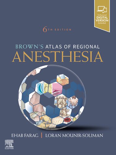
Brown is Atlas of Regional Anesthesia 6th Edition by Ehab Farag, Loran Mounir Soliman 0323654355 9780323654357
$50.00 Original price was: $50.00.$25.00Current price is: $25.00.
Brown is Atlas of Regional Anesthesia 6th Edition by Ehab Farag, Loran Mounir Soliman – Ebook PDF Instant Download/Delivery: 0323654355, 9780323654357
Full download Brown is Atlas of Regional Anesthesia 6th Edition after payment

Product details:
ISBN 10: 0323654355
ISBN 13: 9780323654357
Author: Ehab Farag, Loran Mounir Soliman
Trusted by anesthesiologists, nurse anesthetists, and pain management specialists through five outstanding editions, Brown’s Atlas of Regional Anesthesia, 6th Edition, continues to keep you fully informed and up to date in this fast-changing field. This practical how-to guide demonstrates each technique in an easy-to-follow manner, providing unmatched guidance on administering a wide range of nerve block techniques in all areas of the body. New videos, new illustrations, and new chapters improve your knowledge and expertise, helping you provide optimal, safe regional anesthesia to every patient.
Brown is Atlas of Regional Anesthesia 6th Table of contents:
Section I: Introduction
1. Pharmacology
Drugs
Local anesthetic toxicity
2. Pharmacology of local anesthetics in pediatrics
Introduction
Amide local anesthetics
Ester local anesthetics
Toxicity of local anesthetics
Suggested reading
3. Equipment and ultrasound
Equipment
Section II: Upper Extremity Blocks
4. Upper extremity block anatomy
5. Interscalene block
Perspective
Traditional block technique
Ultrasound for interscalene block
6. Supraclavicular block
Perspective
Traditional block technique
Ultrasonography-guided technique
7. Suprascapular block
Sonoanatomy
Indications
Technique
Pearls
Suprascapular block anterior approach
8. Infraclavicular block
Perspective
Traditional block technique
Potential problems
Ultrasonography-guided technique
Key points
References
9. Axillary block
Perspective
Traditional block technique
Sonoanatomy
Axillary catheter technique
10. Distal upper extremity blocks
Sonoanatomy
Technique
Digital block
Pearls
11. Intravenous regional block
Perspective
Placement
Potential problems
Pearls
Section III: Lower Extremity Blocks
12. Lower extremity block anatomy
13. Lumbar plexus block
Inguinal perivascular block (three-in-one block)
Psoas compartment block
Lumbar plexus block
14. Sciatic block
Perspective
Traditional block technique
Potential problems
Pearls
Ultrasonography-guided technique
15. Femoral block
Perspective
Traditional block technique
Ultrasonography-guided technique
16. Ultrasound for fascia iliaca and inguinal region blocks
Perspective
Anatomy
Sonoanatomy
Inguinal region blocks
Indication
Anatomy
17. Lateral femoral cutaneous nerve block
Anatomy
Landmark technique
Ultrasound-guided technique
Pearls
18. Obturator block
Perspective
Placement
Potential problems
Pearls
Sonoanatomy
Indications
Technique
Pearls
19. Popliteal and saphenous block
Perspective
Traditional block technique
Ultrasonography-guided technique
20. Adductor canal block
Sonoanatomy
Technique
Pearls
21. Ankle block
Perspective
Traditional block technique
Ultrasonography-guided technique
Section IV: Head and Neck Blocks
22. Retrobulbar (peribulbar) block
23. Cervical plexus block
Sonoanatomy
Technique
Pearls
24. Stellate ganglion block
Perspective
Placement
Potential complications
Pearls
Section V: Airway Blocks
25. Airway block anatomy
26. Glossopharyngeal block
Perspective
Placement
Potential problems
Pearls
27. Superior laryngeal block
Perspective
Placement
Potential problems
Pearls
28. Translaryngeal block
Perspective
Placement
Potential problems
Pearls
Section VI: Truncal Blocks
29. Truncal block anatomy
30. PECS and Pecto-Intercostal blocks
Perspective and background
PECS 1 block
PECS 2 block
Pecto-intercostal block
Potential complications
Pearls
31. Serratus anterior block
Relevant anatomy
Potential complications
32. Ultrasound for intercostal block
Sonoanatomy
Technique
Pearls
33. Paravertebral block
Indications
Contraindications
Sonoanatomy
Technique
Pearls
34. Erector spinae plane block
Perspective
Potential complications
Pearls
35. Rectus sheath block and catheter in adults
Sonoanatomy
Technique
Pearls
36. Transversus abdominis plane block (classic approach)
Relevant anatomy
Technique
Indications
Pearls
37. Subcostal transversus abdominal plane block
Sonoanatomy
Technique
Indication
Pearls
38. Quadratus lumborum block
Sonoanatomy
Techniques (fig. 38.3–38.8)
Pearls
Section VII: Neuraxial Blocks
39. Ultrasound-assisted neuraxial blocks
Relevant sonoanatomy of the spine
Technique
Pearls
40. Spinal block
Perspective
Placement
Potential problems
Pearls
41. Epidural block
Perspective
Placement
Potential problems
Pearls
42. Caudal block
Perspective
Placement
Potential problems
Pearls
Section VIII: Pediatric Regional Using Ultrasound
43. Caudal block in pediatrics
Position
Anatomy
Sonoanatomy
Technique
Pearls
44. Ilioinguinal and iliohypogastric block
Position
Anatomy
Sonoanatomy and technique
Pearls
45. Superficial cervical plexus block
Position
Anatomy and technique
Pearls
46. Pudendal nerve block
Indications
Contraindications
Sonoanatomy
Technique
Pearls
47. Paravertebral catheters in pediatrics
Indications
Contraindications
Sonoanatomy
Technique
Pearls
Section IX: Obstetrics Regional Anesthesia
48. Regional techniques during pregnancy and delivery
Perspectives
Patient selection
Pharmacologic choice
Placement
Needle puncture
Potential problems
Pearls
People also search for Brown is Atlas of Regional Anesthesia 6th:
brown’s atlas of regional anesthesia
brown’s atlas of regional anesthesia pdf
atlas of regional anesthesia pdf
atlas of regional anesthesia
Tags:
Ehab Farag,Loran Mounir Soliman,Regional,Anesthesia
You may also like…
Medicine - Anesthesiology and Intensive Care
Medicine - Anatomy and physiology
Netter Atlas of Human Anatomy Classic Regional Approach 8th edition Netter Md
Engineering - Civil & Structural Engineering
Finite Element Analysis and Design of Steel and Steel Concrete Composite Bridges Ehab Ellobody
Housekeeping & Leisure - Games: Chess
Medicine - Anesthesiology and Intensive Care
Romance - Contemporary Romance
Uncategorized
Military Advanced Regional Anesthesia and Analgesia Handbook 2nd Edition Chester Buckenmaier
Housekeeping & Leisure - Games: Chess
Uncategorized
Anesthesiology In Training Exam Review Regional Anesthesia and Chronic Pain Ratan K Banik Editor


