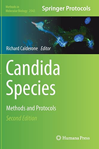
Candida Species Methods and Protocols 2nd Edition by Richard Calderone ISBN 9781071625484 1071625489
$50.00 Original price was: $50.00.$25.00Current price is: $25.00.
Candida Species Methods and Protocols 2nd Edition by Richard Calderone – Ebook PDF Instant Download/Delivery: 9781071625484 ,1071625489
Full download Candida Species Methods and Protocols 2nd Edition after payment
Product details:
ISBN 10: 1071625489
ISBN 13: 9781071625484
Author: Richard Calderone
This updated volume explores Candida and candidiasis methods, useful to a wide variety of Candida scientists including those new to the field. Beginning with a section on the Candida genome, the book continues by covering techniques for antifungal discovery and studying resistance, pathogenesis and virulence, communities, as well as immunity. Biofilm protocols are also featured. Written for the highly successful Methods in Molecular Biology series, chapters include introductions to their respective topics, lists of the necessary materials and reagents, step-by-step, readily reproducible laboratory protocols, and tips on troubleshooting and avoiding known pitfalls.
Authoritative and up-to-date, Candida Species: Methods and Protocols, Second Edition serves as an ideal guide for researchers working to further our understanding of this family of infective fungi.
Candida Species Methods and Protocols 2nd Edition Table of contents:
Part I: The Candida Genome
Chapter 1: CRISPR-Mediated Genome Editing in the Human Fungal Pathogen C. albicans
1 Introduction
2 Materials
2.1 Enzymes and Buffers
2.2 Reagents: DNA Extraction
2.3 Media for Growth of C. albicans
3 Methods
3.1 Design and Anneal Guide Oligonucleotides
3.2 Clone Guide Sequence Into pV1393 and Transform Into Escherichia coli DH5α
3.3 Design and Prepare Repair Templates
3.4 C. albicans Transformation
3.5 Colony PCR and Screening
3.6 Removal of CRISPR Cassette
4 Notes
References
Chapter 2: Plasmid-Based CRISPR-Cas9 Editing in Multiple Candida Species
1 Introduction
2 Materials
2.1 Media and Strains
2.2 Plasmid Preparation and Cloning
2.3 Chemical Transformation of Candida parapsilosis sensu lato
2.4 Transformation of Candida tropicalis Using Electroporation
3 Methods
3.1 At the Desk: Design of the Strategy
3.1.1 Selecting a Guide (Protospacer) Targeting the Gene of Interest (YFG)
3.1.2 Designing a Repair Template (RT) to Introduce Premature Stop Codons
3.1.3 Designing a Repair Template for Gene Deletion (RTdel-YFG)
3.1.4 Designing Oligonucleotide Primers for Screening
3.2 At the Bench: Cloning, RT Synthesis, and Transformation of Candida
3.2.1 Generate pCP-YFG or pCT-YFG
3.2.2 pCP-tRNA-YFG and pCT-tRNA-YFG Plasmid Extraction
3.2.3 Synthesize the Repair Template (RT): C. parapsilosis
3.2.4 Synthesize the Repair Template (RT): C. tropicalis
3.2.5 Chemical Transformation in Candida parapsilosis
3.2.6 Electroporation in Candida tropicalis
3.2.7 Screening of Candida Transformants and Plasmid Curing
4 Notes
References
Chapter 3: A Proteomic Approach for the Quantification of Posttranslational Protein Lysine Acetylation in Candida albicans
1 Introduction
2 Materials
2.1 Chemicals and Reagents
2.2 Buffers and Solutions
3 Methods
3.1 Cell Lysis
3.2 Protein Precipitation
3.3 Protein Quantification
3.4 Tryptic Digestion (In-Solution Digestion)
3.5 Desalting and Lyophilization Using Sep-Pak tC18 Solid-Phase Extraction
3.6 TMT Labeling
3.7 Quality Check for Labeling Efficiency
3.8 Pool the TMT-Labeled Samples
3.9 Fractionation by Reversed-Phase HPLC
3.9.1 Pool the Peptide Fractions
3.10 Ac-K Enrichment
3.11 C18 Stage-Tipping
3.12 LC-MS/MS Analysis
3.13 MS Data Processing and Analysis
4 Notes
References
Chapter 4: How to Use the Candida Genome Database
1 Introduction
2 Methods
3 GO Term Finder and GO Slim Mapper, Tools for GO Analysis
3.1 Using GO Term Finder
3.2 Using GO Slim Mapper
4 Analyze the Genome Using JBrowse
5 Using the Advanced Search and Batch Download Tools
5.1 Using the Advanced Search Tool
5.2 Using the Batch Download Tool
6 Notes
References
Chapter 5: Subtelomeric Chromatin Structure by Chromosome Conformation Capture (3C)-qPCR Methodology in Candida glabrata
1 Introduction
2 Materials
2.1 Reagents for 3C DNA Cross-Linking
2.2 Reagents for 3C Libraries
2.3 Reagents for DNA Concentration, 3C-qPCR
2.4 Reagents for Random Ligation Protocol
3 Methods
3.1 Choice of Restriction Enzymes
3.2 Design of 3C Test Primers, Anchor Primer, and TaqMan Probe
3.3 Cell Growth
3.4 Formaldehyde Cross-Linking of Chromatin
3.5 Digestion
3.6 Ligation of Cross-Linked Fragments
3.7 Ligation Products Purification
3.8 Loading Adjustments
3.9 3C-qPCR
3.10 Random Ligation
3.11 Determination of Digestion Efficiency
3.12 Data Analysis
4 Notes
References
Chapter 6: Construction of Double Heterozygous Deletion Strains for Complex Haploinsufficiency-Based Genetic Analysis in Candi…
1 Introduction
2 Materials
2.1 Transcription Factor Deletion Cassette
2.2 Transformation Reagents
2.3 Confirm Heterozygous Deletion of Genes
2.4 Sample CHI Analysis Process: Biofilm Assay and Calculation of Epsilon Genetic Interaction Scores
3 Methods
3.1 Generation of the Transcription Factor Deletion Cassette
3.2 Preparation of Competent Yeast for Transformation
3.3 Transformation Reaction
3.4 Heat Shock and Plating Yeast
3.5 Confirm Heterozygous Deletion of Genes
3.6 Example of the CHI Analysis Process: Biofilm Assay and Calculation of Epsilon Genetic Interaction Scores
4 Notes
References
Part II: Antifungal Discovery and Resistance
Chapter 7: Using Synthetic Genetic Interactions in Candida glabrata as a Novel Method to Detect Genes with Roles in Antifungal…
1 Introduction
2 Materials and Methods
2.1 Yeast Strains
2.2 Cloning Strategy for Generation of Query Strain
2.3 Agar Plates for Synthetic Genetic Interaction Screen
2.4 Genetic Interaction Screen
2.5 Data Analysis
2.6 Network Mapping and Confirmation
2.7 Conclusion
3 Notes
3.1 Defining an Interaction
References
Chapter 8: Identifying Specific Small Molecule-Protein Interactions Using Target Abundance-Based Fitness Screening (TAFiS)
1 Introduction
2 Materials
3 Methods
3.1 Construction of Candida albicans Strains with Differential Target Protein Expression
3.2 High-Throughput Chemical Screening Assay
3.3 Dose-Response Confirmation of Primary Hits
3.4 Counter-Screen to Eliminate False Positives
4 Notes
References
Chapter 9: High-Throughput Phospholipidomics of Candida Cells: From Sample Preparation to Data Analysis
1 Introduction
2 Materials
2.1 Candida Strains
2.2 Cell Culture
2.3 Chemicals
2.4 Equipment
3 Methods
3.1 Culture Conditions
3.2 Lipid Extraction
3.2.1 Folch Method
3.2.2 Bligh and Dyer Extraction
3.3 Protein Estimation (BCA Assay)
3.4 Buffers and HPLC Separation
3.5 Mass Spectrometry (MS) Analysis
3.6 Data Analysis
3.6.1 Lipid Quantification
3.6.2 Data Normalization
4 Notes
References
Chapter 10: Genetic Screens of an Anti-Candida Natural Product Using the Heterozygous Saccharomyces cerevisiae Mutant Library
1 Introduction
2 Materials
2.1 Strains and Reagents
2.2 Equipment
3 Methods
3.1 Determination of Minimum Inhibitory Concentration (MIC) and Minimum Fungicide Concentration (MFC) of Natural Products
3.2 Screening the MOA with Heterozygous Mutant Library
3.2.1 Procedure for Sterilizing the Pin Replicator (See Note 5)
3.2.2 Replication of the Collection (See Note 6)
3.2.3 Preliminary Experiment to Determine an Appropriate Concentration of Natural Product for Fitness Profiling Experiments (S…
3.2.4 Screening for Hypersensitive Mutants with the Natural Product
4 Notes
References
Chapter 11: Assessing Complex I (CI) Mitochondrial Subunit Protein Functions in Candida albicans
1 Introduction
2 Materials
2.1 Strains of C. albicans and Growth Media
2.2 Drop-Plate Assays; ROS Compounds
2.3 ROS Measurements
2.4 Oxygen Uptake by Fungal Cells
2.5 Chronological Life Span (CLS)
2.6 Mitochondrial Membrane Potential
2.7 Spheroplast Preparation
2.8 Isolation of Mitochondria
2.9 Mitochondria-Enriched Isolation
2.10 Extraction of Semi-purified Respiratory Complex Proteins CI-CIV
2.11 Blue Native (BN) Polyacrylamide Gel Electrophoresis of CI-CIV Respiratory Complexes
2.12 Enzyme Assays of CI-CIV Proteins (Performed with 20 μg of Semi-purified Mitochondrial Proteins)
2.13 Visualizing Mitochondria
2.14 TCA Cycle and Associated Enzymes
2.15 The Energy Phenotypes of Wild Type and Mutants in ETC CI, III, and CIV. The XF96 Seahorse Assays
3 Methods
3.1 Strains and Their Growth
3.2 Drop Plate Assays
3.3 Reactive Oxidant Species Measurements (ROS)
3.4 Mitochondria Membrane Potential
3.5 Oxygen Uptake
3.6 Mitochondria Isolation
3.7 Assays of Mitochondria ETC CI-IV Enzyme Activity
3.8 TCA and Accessory Enzyme Assays
4 Notes
References
Part III: Pathogenesis and Virulence
Chapter 12: In Vitro Biophysical Characterization of Candidalysin: A Fungal Peptide Toxin
1 Introduction
2 Materials
2.1 Candidalysin 3 mM Stock Preparation
2.2 Candidalysin 300 μM Stock for Electrical Recording
2.3 Candidalysin 500-μM Stock for Far-UV CD Experiments
2.4 Lipid Stock Solution
2.5 Detergent Stock Solution
2.6 1-M HEPES Stock Buffer
2.7 3-M Potassium Chloride (KCl) Stock Buffer
2.8 Orbit Measuring Buffer: 20-mM HEPES pH 7.4, 100-mM KCl
2.9 CD Measuring Buffer: 0.1-M Tris-HCl pH 7.4
2.10 Microelectrode Cavity Array (MECA) Chip Cleaning Reagents
2.11 Quartz Cuvette Cleaning Reagents
3 Methods
3.1 Orbit e16 Instrument Preparation and Parameter Settings
3.2 Calibrating Orbit e16
3.3 Orbit e16 Assembly
3.4 Preparation of Horizontal Lipid Bilayers
3.5 Measuring the Real-Time Permeabilization by Candidalysin
3.6 CD Instrument Preparation and Parameter Settings
3.7 Sample Preparation for CD Experiments
3.8 CD Spectrum Acquisition
3.9 Data Processing and Analysis of Secondary Structure by CD Spectroscopy
4 Notes
References
Chapter 13: Assessment of cAMP-PKA Signaling in Candida glabrata by FRET-Based Biosensors
1 Introduction
2 Materials
2.1 Plasmids and Materials for Generation of Strains
2.2 Growth Media and Chemicals for Sample Preparation
2.3 Cell Immobilization
2.4 Fluorescence Microscopy and Attributes
3 Methods
3.1 Transformation of Candida glabrata
3.2 Determining the Settings for Fluorescence Microscopy and FRET
3.3 Growing Strains for FRET Measurement
3.4 Microscope Handling and Settings
3.5 Preparation of the Immobilized Cells Using a Microfluidic Device or Manual Mode
3.5.1 Microfluidic Settings
3.5.2 Preparation of the Microfluidic System
3.5.3 Preparation of the Immobilized C. glabrata Cells Using Concanavalin A
3.6 Data Acquisition
3.7 Data Analysis and Representation
3.7.1 ROI Selection
3.7.2 FRET Calculation
AKAR3-PKA Activity Calculation
EPAC2-cAMP Level Calculation
4 Notes
References
Chapter 14: Methods Related to the Immunopathogenesis of Vulvovaginal Candidiasis and Associated Neutrophil Anergy
1 Introduction
2 Methodology
2.1 Principal Protocol: A Mouse Model of Acute/Chronic VVC
2.1.1 Materials
2.1.2 Methods
2.1.3 Notes
3 Support Protocols: Outcome Measures for Vaginitis in Mice
3.1 Vaginal Fungal Burden
3.1.1 Materials
3.1.2 Methods
3.1.3 Notes
3.2 Wet Mount Microscopy
3.2.1 Materials
3.2.2 Methods
3.2.3 Notes
3.3 Vaginal Secretions
3.3.1 Materials
3.3.2 Methods
3.3.3 Notes
3.4 Cytological/Histological Examinations: Papanicolaou Stain
3.4.1 Materials
3.4.2 Methods
3.4.3 Notes
3.5 Assessment of NETotic Activity in In Vitro and In Vivo Models
3.5.1 Materials
3.5.2 Methods
3.5.3 Notes
4 Additional Methods for Use of Vaginal Tissues
4.1 Vaginal Tissue Resection
4.1.1 Materials
4.1.2 Methods
4.1.3 Notes
4.2 Use of Vaginal Explants in an Ex Vivo Model of Vaginal C. albicans Infection/Biofilm Formation
4.2.1 Materials
4.2.2 Methods
4.2.3 Notes
4.3 Establishment of Primary Vaginal Epithelial Cell Cultures
4.3.1 Materials
4.3.2 Methods
5 Methods Associated with Administration of Drugs for Treatment of VVC
5.1 Materials
5.2 Methods
5.3 Notes
6 Assays Related to “Neutrophil Anergy ́ ́
6.1 PMN Killing Assay Under Vaginal Simulation Conditions
6.1.1 Preparation of Vaginal Conditioned Media (VCM)
6.1.2 Isolation of Elicited Peritoneal PMNs
6.1.3 Isolation of Elicited Vaginal PMNs
6.1.4 PMN Killing Assay
6.2 Assays Testing for the Presence of Heparan Sulfate (HS)
6.2.1 Materials
6.2.2 Methods
6.2.3 Notes
7 Missing Pieces/Unanswered Questions/Future Studies
8 Conclusions
References
Chapter 15: A Simple Method for Growth of Candida albicans Biofilms Under Continuous Media Flow and for Recovery of Biofilm Di…
1 Introduction
2 Materials
2.1 Fungal Isolates
2.2 Reagents
2.3 Equipment and Materials
2.4 Reagent Setup
2.5 Equipment Setup
3 Methods
3.1 Adhesion Step
3.2 Biofilm Formation
3.3 Expected Results
4 Notes
References
Chapter 16: A Simple 96-Well Plate-Based Method for Development of Candida Biofilms Under Static Conditions
1 Introduction
2 Experimental Design
2.1 Materials
2.1.1 Fungal Isolates
2.1.2 Reagents
2.1.3 Acetone
2.1.4 Hemacytometer
2.1.5 Inverted Microscope
2.1.6 Microtiter Plate Reader
2.1.7 Reagents Setup
2.2 Methods
2.2.1 Growth of Fungal Cultures and Preparation of Inocula
2.2.2 Biofilm Formation
2.2.3 Antifungal Challenge of Pre-formed Biofilms
2.2.4 Post-processing and Measurement of Metabolic Activity After Antifungal Treatment
2.2.5 Expected Results
3 Notes
References
Chapter 17: Comparative Analysis of the Fitness of Candida albicans Strains During Colonization of the Mice Gastrointestinal T…
1 Introduction
2 Materials
3 Methods
3.1 Experimental Design
3.2 Mice Preparation and Candida Inoculation
3.3 Tracing Viable Fungal Populations
3.4 Prompt Estimation of Fungal Population
3.5 Determination of Fungal Loads within the Gastrointestinal Tract
3.6 Representation and Analysis
4 Notes
References
Chapter 18: Detection and Identification of Candida auris from Clinical Skin Swabs
1 Introduction
2 Materials
2.1 Patient Swabs
2.2 DNA Extraction from Swab Medium
2.3 Candida auris-Specific Real-Time PCR with an Internal Amplification Control (See Note 4)
2.4 Candida auris Enrichment Broth
2.5 DNA Extraction from Colonies
2.6 Amplification of rDNA
2.7 Gel Electrophoresis
2.8 Sanger Sequencing of ITS PCR Products and Sequence Analysis
3 Methods
3.1 DNA Extraction from Patient Swabs
3.2 Candida auris-Specific Real-Time PCR Assay with an Internal Amplification Control
3.3 Isolation of Candida auris Using Salt Sabouraud Dulcitol Enrichment Broth
4 Notes
References
Chapter 19: Production and Isolation of the Candida Species Biofilm Extracellular Matrix
1 Introduction
2 Materials
2.1 Growth Media for Small- and Large-Scale Matrix Production
2.2 Reagents for Large-Scale Matrix Production
3 Methods
3.1 Small-Scale Production and Isolation of Biofilm Matrix (6-Well Method)
3.1.1 Inoculum Preparation and Biofilm Seeding of 6-Well Culture Plates
3.1.2 Harvesting of Biofilms
3.2 Large-Scale Production and Isolation of Biofilm Matrix (Roller Bottle System)
3.2.1 Inoculum Preparation and Biofilm Seeding of Roller Bottles
3.2.2 Harvesting of Biofilms
3.2.3 Matrix Dialysis
4 Notes
References
Part IV: Communities
Chapter 20: Characterization of the Effects of Candida Gastrointestinal Colonization on Clostridioides difficile Infection in …
1 Introduction
2 Materials
2.1 Animal Models
2.2 C. difficile Spore Preparation
2.3 Measuring Toxin Activity
3 Methods
3.1 Animal Models
3.2 C. difficile Spore Preparation
3.3 Measuring Toxin Activity
4 Notes
References
Chapter 21: Analysis of Gut Microbiome Using Fecal Samples
1 Introduction
2 Materials
2.1 Sample Collection/Processing
2.2 DNA Processing
2.3 PCR Amplification
2.4 Library Preparation and Sequencing
3 Methods
3.1 Fecal Swab Processing
3.2 DNA Extraction
3.3 PCR Amplification
3.4 Library Preparation and Sequencing
3.4.1 End Repair
3.4.2 Ligation
3.4.3 Library Pooling
3.4.4 Size Selection Using the Pippin Preparation (Optional Step)
3.4.5 Quantitative PCR
3.4.6 Chef
3.4.7 Sequencer
3.5 Statistical Analysis
4 Notes
Appendix A
References
Untitled
Chapter 22: Measurements of Treg Cell Induction by Candida albicans DNA Using Flow Cytometry
1 Introduction
2 Materials
2.1 Reagents and Antibodies Used for Fluorescence-Activated Cell Sorting (FACS)
3 Methods
3.1 Fungal Cell Growth, Genomic DNA (gDNA) Extraction and Purification
3.1.1 Enzymatic Method for gDNA Preparation by Blood and Cell Culture DNA Mini Kit
3.1.2 Method for gDNA Preparation
3.2 Isolation of CD4+ T Cells from PBMC
3.3 In Vitro Induction of Treg by Microbial Genomic DNA
3.4 Cell Staining and FACS Analysis
4 Notes
References
Part V: Immunity
Chapter 23: Molecular and Microscopic Methods of Quantifying Candida albicans Cell Wall PAMP Exposure
1 Introduction
2 Materials
2.1 Materials for Candida Growth
2.2 Staining Reagents
2.3 Other Required Reagents
3 Methods
3.1 Standard Reagent Preparation
3.1.1 Yeast Peptone Dextrose (YPD)
3.2 Prepare C. albicans Cells
3.3 Staining of C. albicans Cell Wall Components
3.3.1 Fc-Dectin-1 Staining
3.3.2 Mannan and Exposed Chitin Staining
3.3.3 Staining Total Chitin and Glucan
3.4 Quantitative Analysis by Flow Cytometry
3.5 Analysis by Fluorescent Microscopy
3.6 Alcian Blue Staining
4 Data Analysis (Sample)
4.1 Flow Cytometry
4.2 Fluorescent Microscopy
5 Summary
6 Notes
References
Chapter 24: Isolation, Physicochemical Characterization, Labeling, and Biological Evaluation of Mannans and Glucans
1 Introduction
2 Materials
2.1 Growth of C. albicans for Mannan/Glucan Extraction
2.2 Isolation of Mannans
2.3 Isolation of Glucans
2.4 Molecular Weight Determination of Mannans and Glucans
2.5 NMR Structural Analysis of Mannans
2.6 NMR Structural Analysis of Glucans
2.7 Depyrogenation and Sterilization of Mannans
2.8 Depyrogenation and Sterilization of Water-Insoluble Glucan Particles
2.9 Functionalization and Labeling of Mannans and Glucans
2.10 Characterizing the Binding Interactions of Water-Soluble Mannans and Glucans with Human Innate Immune Pattern Recognition…
2.11 Assessment of Dectin-1 Binding to Particulate Glucan
2.12 Sterilization of the Glucan Particle Suspension
2.13 Endotoxin Testing of the Glucan Particle Suspension
2.14 Assay for In Vitro Bioactivity of Mannans and Glucans
3 Methods
3.1 Growth of C. albicans for Mannan/Glucan Extraction
3.2 Isolation of Mannans
3.3 Isolation of Glucans
3.3.1 Base (NaOH) Extraction
3.3.2 Acid (H3PO4) Extraction
3.3.3 Acid/Ethanol Extraction (See Note 30)
3.3.4 Water Wash
3.3.5 Interim 1H-NMR Analysis (See Note 31)
3.3.6 Amylase Treatment of C. albicans Glucan to Remove Residual Glycogen (see Note 32)
3.3.7 Pronase Treatment to Remove Residual Protein (See Note 34)
3.4 Molecular Weight Determination of Water-Soluble Mannans and Glucans
3.5 NMR Structural Analysis of Mannans
3.6 NMR Structural Analysis of Glucans
3.7 Depyrogenation and Sterilization of Water-Soluble Mannans and Glucans
3.8 Depyrogenation and Sterilization of Water-Insoluble Glucan Particles (See Note 60)
3.9 Functionalization and Labeling of Mannans and Glucans
3.9.1 DAP Derivatization for Water-Insoluble Glucan Particles
3.9.2 DAP Derivatization for Water-Soluble Glucans or Mannans
3.9.3 Conjugation of DAP Derivatized Carbohydrate with Fluorescent Dyes
3.10 Characterizing the Binding Interactions of Water-Soluble Mannans and Glucans with Human Innate Immune Pattern Recognition…
3.11 Assessment of Dectin-1 Binding to Particulate Glucan (See Note 81)
3.12 Sterilization of a Glucan Particle Suspension (See Note 82)
3.13 Endotoxin Testing of the Glucan Particle Suspension
3.13.1 This Assay Will Determine Whether the Glucan Particle Suspension Contains Detectable Endotoxin
3.13.2 Final QA/QC 1H-NMR Analysis of Glucans (See Note 87)
3.13.3 Sterility Test of the Glucan Particle Suspension
Testing for Bacterial Contamination by Culture
Testing for Mycoplasma Contamination
3.14 Bioactivity of Mannans and Glucans
4 Notes
References
Chapter 25: Understanding the Protective Role of IL-17 During Oropharyngeal Candidiasis
1 Introduction
2 Materials
2.1 YPD Agar and Broth Preparation (Day -3)
2.2 Cortisone Acetate Preparation and Administration (Day -1)
2.3 Anesthetic and Saline Preparation (Day -1)
3 Methods
3.1 Overnight Yeast Culture (Day -1)
3.2 Yeast Suspension Preparation (Day 0, Day of Infection)
3.3 Candida albicans Infection (Day 0) (See Note 5)
3.4 Harvest Preparation (Day 4)
3.5 Sacrifice and Tissue Harvest (Day 5)
3.6 Plating Tissue Homogenate (Day 5)
4 Support Protocol 1: Hematoxylin and Eosin Staining of Tongue Sections
4.1 Materials
4.2 Procedure
5 Support Protocol 2: Periodic Acid-Schiff Staining
5.1 Materials
5.2 Procedure
6 Support Protocol 3: Tongue Tissue Processing for Flow Cytometric Analysis
6.1 Materials
6.2 Procedure
7 Support Protocol 4: Salivary Collection and Candidacidal Assay (See Note 12)
7.1 Materials
7.2 Procedure
8 Notes
References
Index
People also search for Candida Species Methods and Protocols 2nd Edition:
is candida species contagious
candida species swab
candida species – wet mount
candida species vs candida glabrata
candida species tma
You may also like…
Uncategorized
Candida auris Methods and Protocols Methods in Molecular Biology 2517 Alexander Lorenz (Editor)
Medicine - Pharmacology
Antibiotics Methods and Protocols 2nd Edition by Peter Sass 1071628577 978-1071628577
Biology and other natural sciences - Molecular
Biology and other natural sciences - Biology
Peroxisomes Methods and Protocols 2nd Edition by Michael Schrader ISBN 9781071630471 1071630474
Uncategorized
Chaperones Methods and Protocols 2nd Edition by John M Walker ISBN 9781071633427 1071633414

