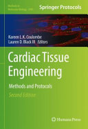
Cardiac Tissue Engineering 2nd Edition by Kareen LK Coulombe, Lauren D Black III ISBN 9781071622605 1071622609
$50.00 Original price was: $50.00.$25.00Current price is: $25.00.
Cardiac Tissue Engineering 2nd Edition by Kareen LK Coulombe, Lauren D Black III – Ebook PDF Instant Download/Delivery: 9781071622605 ,1071622609
Full download Cardiac Tissue Engineering 2nd Edition after payment
Product details:
ISBN 10: 1071622609
ISBN 13: 9781071622605
Author: Kareen LK Coulombe, Lauren D Black III
This detailed volume presents an updated collection of state-of-the-art protocols in cardiac tissue engineering. These protocols demonstrate advancements in cell sourcing, assembly, and use of engineered cardiac tissues, imaging and diagnostics, as well as therapeutic applications. New animal models, biomaterials, and quantitative analyses are described for broad adoption. Written for the highly successful Methods in Molecular Biology series, chapters include introductions to their respective topics, lists of the necessary materials and reagents, step-by-step, readily reproducible laboratory protocols, and tips on troubleshooting and avoiding known pitfalls.
Authoritative and practical, Cardiac Tissue Engineering: Methods and Protocols, Second Edition serves as an ideal resource for inspiring the advancement of cardiotoxicity assessment, drug discovery, and heart repair and regeneration in order to accelerate heart health improvement around the globe
Cardiac Tissue Engineering 2nd Edition Table of contents:
Chapter 1: CRISPR Library Screening in Cultured Cardiomyocytes
1 Introduction
2 Materials
2.1 Molecular Biology
2.2 Cell Culture
3 Methods
3.1 Library Preparation and In Silico sgRNA Sequence Design
3.2 Sequence Design for sgRNA Oligonucleotide Cloning
3.3 sgRNA Library Plasmid Cloning
3.4 Assessment of Baseline Cardiomyocyte Proliferation
3.5 Assessment of Antibiotic Susceptibility
3.6 sgRNA Lentivirus Library Preparation
3.7 Library Delivery and Screening
3.8 Validation of sgRNA Targets
4 Notes
References
Chapter 2: Protein and mRNA Quantification in Small Samples of Human-Induced Pluripotent Stem Cell-Derived Cardiomyocytes in 9…
1 Introduction
2 Materials
2.1 hiPSC-CMs Cell Culture
2.2 Cell Lysis and Protein Denaturing for WB
2.3 Immunoprobing
2.4 qPCR with the Cells-to-CT Kit
3 Methods
3.1 Culturing hiPSC-CMs in a 96-Well Plate (50,000 Cells per Well)
3.2 Adenovirus Infection-Optional to Illustrate Usage
3.3 Protein Collection from a 96-Well Microplate
3.4 Protein Quantification Using Wes
3.4.1 Experiment Planning and Setting the Compass Software
3.4.2 Sample and Primary Antibody Preparation for Protein Quantification
3.4.3 Microplate Loading and Start of a Wes Run
3.4.4 Data Analysis of Wes Data
3.5 Primary Antibody Dilution Optimization
3.6 Normalized Protein Expression by Antibody Multiplexing and Total Protein Linear Response Test
3.7 mRNA Quantification in hiPSC-CMs Using Cells-to-CT Kit and qPCR
3.8 Example Application
4 Notes
References
Chapter 3: Self-Assembled Heterotypic Cardiac Spheroids from Human Pluripotent Stem Cells
1 Introduction
2 Materials
3 Methods
3.1 Replica Molding to Fabricate Sheets of PDMS Microwell Molds
3.2 Repeat Replica Molding to Create Arrayed Circular Inverse Molds
3.3 Casting Agarose Microwell Molds Off of PDMS-Arrayed Circular Inverse Molds
3.4 Cell Seeding into Agarose Microwell Molds
3.5 Removing Microtissues from Agarose Microwell Molds
3.6 Rotary Suspension Culture
4 Notes
References
Chapter 4: Acellular Myocardial Scaffolds and Slices Fabrication, and Method for Applying Mechanical and Electrical Simulation…
1 Introduction
2 Materials
2.1 Decellularization Stock Solution
2.2 Porcine Myocardium
2.3 Cell Culture Medium, Differentiation Medium, and Complete Medium
3 Methods
3.1 Preparation and Handling Tips for Acellular Myocardial Slices
3.2 Preparation of the Multi-stimulation Bioreactor
3.3 Bioreactor Setup and Sterilization for Acellular Tissue Construct
3.4 MSC Culture, Reseeding, Differentiation, and Bioreactor Conditioning Protocol
4 Notes
References
Chapter 5: FRESH 3D Bioprinting a Ventricle-like Cardiac Construct Using Human Stem Cell-Derived Cardiomyocytes
1 Introduction
2 Materials
3 Methods
3.1 Prepare Bioprinting Reagents
3.2 Design Cardiac Ventricle Model
3.3 Preparation of Cellular Ink
3.4 Setup of Bioprinter for Dual Extrusion Fabrication
3.5 Alignment of Extruder Needle Tips
3.6 Preparation of FRESH Support Material
3.7 Setup of Printer Stage and Print Initiation
3.8 Release and Culture of Ventricle Model
4 Notes
References
Chapter 6: Engineered Heart Tissues for Contractile, Structural, and Transcriptional Assessment of Human Pluripotent Stem Cell…
1 Introduction
2 Materials
2.1 PDMS Post Racks and PLA Spacer Arrays
2.2 Cell Culture
2.3 EHT Casting
2.4 EHT Analysis
3 Methods
3.1 PDMS Post Racks and Preparation Prior to Casting
3.2 Cell Preparation
3.3 Agarose Wells, Thrombin Aliquots, and Fibrinogen
3.4 EHT Preparation
3.5 EHT Casting
3.6 EHT Culture
3.7 EHT Analysis
3.7.1 Optical Force Measurement
3.7.2 Calcium Handling Assessment
3.7.3 Gene Expression Analysis
3.7.4 Histology and Immunocytochemistry
4 Notes
References
Chapter 7: High-Throughput Analysis of Drug Safety Responses in Induced Pluripotent Stem Cell-Derived Cardiomyocytes Using Mul…
1 Introduction
2 Materials
2.1 Cell Culture
2.2 MEA Experiment
3 Methods
3.1 Cell Plating
3.2 MEA Recording and Acute Drug Testing
3.3 MEA Analysis
4 Notes
References
Chapter 8: iPSC-Derived Micro-Heart Muscle for Medium-Throughput Pharmacology and Pharmacogenomic Studies
1 Introduction
2 Materials
2.1 Stem Cell Culture
2.2 Cardiac Differentiation and Maintenance
2.3 Stencil Manufacturing and Seeding
2.4 Western Blot
2.5 qPCR
3 Methods
3.1 Matrigel Aliquoting
3.2 iPSC Thawing
3.3 iPSC Passaging and Maintenance
3.4 iPSC Freezing
3.5 Cardiomyocyte Differentiation
3.6 Cardiomyocyte Dissociation and Freezing
3.7 Cardiomyocyte Thawing
3.8 Cardiomyocyte Lactate Purification
3.9 3D-Printed Stencil Mold Design
3.10 PDMS Stencil Manufacturing
3.11 Stencil Seeding
3.12 Pharmacology Treatments
3.13 Imaging-Based Physiology Measurements
3.14 Image Analysis
3.15 Lysing Tissues for qPCR/Western Blot
3.16 Tissue Fixation and OCT Embedding
3.17 Tissue Sectioning
3.18 Slide Staining
4 Notes
References
Chapter 9: Quantifying Propagation Velocity from Engineered Cardiac Tissues with High-Speed Fluorescence Microscopy and Automa…
1 Introduction
2 Materials
2.1 Live Imaging of Calcium Wave Propagation in Engineered Cardiac Tissues with Fluo-4
2.2 Calculating Propagation Velocity
3 Methods
3.1 Live Imaging of Calcium Wave Propagation in Engineered Cardiac Tissues with Fluo-4
3.2 Calculating Propagation Velocity
4 Notes
References
Chapter 10: Arrhythmia Assessment in Heterotypic Human Cardiac Myocyte-Fibroblast Microtissues
1 Introduction
2 Materials
2.1 Cardiomyocyte Differentiation
2.2 Fabrication of Hydrogels and 3D Culture
2.3 Optical Mapping and Action Potential Analysis
3 Methods
3.1 Cardiomyocyte Differentiation and Cardiac Fibroblast Maintenance
3.2 Fabrication of Hydrogels and 3D Culture
3.3 Optical Mapping and Automated Action Potential Analysis
4 Notes
References
Chapter 11: Human-Engineered Atrial Tissue for Studying Atrial Fibrillation
1 Introduction
2 Materials
3 Methods
3.1 Preparations
3.2 Differentiation of Atrial-Like hiPSC-Derived Cardiomyocytes (Optional)
3.3 Thawing of hiPSC-Derived Cardiomyocytes (Optional)
3.4 Casting Molds and Preparation of EHT Master Mix (All Steps Carried Out in a Biosafety Cabinet, Fig. 1)
3.5 Generation of Engineered Heart Tissue
3.6 Maintenance
3.7 Video-Optical Contractility Analysis Using EHT Technologies Equipment (Fig. 3)
4 Notes
References
Chapter 12: Design and Fabrication of Biological Wires for Cardiac Fibrosis Disease Modeling
1 Introduction
2 Materials
2.1 SU-8 Master Molds
2.2 PDMS Molds
2.3 POMaC Prepolymer Solution
2.4 Polystyrene Chips
2.5 Cells and Cell Culture Media
2.6 Fibrin-based hydrogel
2.7 Electrical Stimulation Chamber and External Electrical Stimulator
3 Methods
3.1 Part I. Platform Design and Fabrication: Photomask Design and Preparation
3.2 SU-8 Master Mold and PDMS Mold Fabrication
3.3 Polystyrene Chip Fabrication
3.4 POMaC Wire Fabrication and Assembly
3.5 Part II. Tissue Construction and Assessment: Generation of Interstitial Fibrotic Cardiac Tissues and Healthy Controls
3.6 Generation of Focal Fibrotic Cardiac Tissues
3.7 Fabrication of Electrical Stimulation Chambers
3.8 Electrical Stimulation
3.9 Tissue Compaction Assessment
3.10 Polymer Wire Force-Displacement Curves
3.11 Functional Assessment and Force Measurement
3.12 MATLAB Data Analysis
4 Notes
References
Chapter 13: Methods for Transepicardial Cell Transplantation in a Swine Myocardial Infarction Model
1 Introduction
2 Materials
2.1 Induction of Myocardial Infarction
2.2 Implantation of Indwelling VAP
2.3 Thoracotomy, Transepicardial Cell Injection, and Implantation of Subcutaneous Telemetry Device
3 Methods
3.1 Induction of Myocardial Infarction
3.1.1 Pre-anesthesia, General Anesthesia, and Preparation for MI Procedure
3.1.2 MI Induction via Balloon Occlusion of the Mid-LAD
3.2 Implantation of Indwelling VAP
3.2.1 VAP Implantation Procedure
3.2.2 VAP Maintenance and Use
3.3 Thoracotomy, Transepicardial Cell Injection, and Implantation of Subcutaneous Telemetry Device
3.3.1 Pre-anesthesia, General Anesthesia, and Preparation of Surgical Site for Thoracotomy Procedure
3.3.2 Left Lateral Thoracotomy and Cell Implantation Procedure
3.3.3 Implantation of Subcutaneous Telemetry Device
4 Notes
References
Chapter 14: Defined Engineered Human Myocardium for Disease Modeling, Drug Screening, and Heart Repair
1 Introduction
2 Materials
2.1 Cell Culture Components
2.2 EHM Generation Components
3 Methods
3.1 Preparation of EHM Reconstitution and Culture Media
3.2 Preparation of Cells for EHM Generation
3.3 EHM Generation
3.4 Analysis (Quality Control) by Video-Optic Recordings
3.5 Analysis (Quality Control) by Isometric Force Measurements
4 Notes
References
Chapter 15: Tubular Cardiac Tissue Bioengineered from Multi-Layered Cell Sheets for Use in the Treatment of Heart Failure
1 Introduction
2 Materials
2.1 Cell Culture
2.2 Vascular bed
2.3 Tissue Perfusion Bioreactor
2.4 Tubularization of Cardiac Tissue
2.5 Perfusion Medium
2.6 Measurement of Electric Potential and Internal Pressure
2.7 Histological Analysis
3 Methods
3.1 Bioreactor Set-Up
3.2 Fabrication of the Vascular Bed
3.3 Fabrication of Tubular Cardiac Tissue
3.4 Perfusion Culture of Tubular Cardiac Tissue
3.5 Measurements of Electric Potential and Internal Pressure
3.6 Histological Analysis
4 Notes
References
Chapter 16: Quantifying Cardiomyocyte Proliferation and Nucleation to Assess Mammalian Cardiac Regeneration
1 Introduction
2 Materials
2.1 Heart Isolation
2.2 Heart Sectioning
2.3 Immunohistochemistry
2.4 Masson ́s Aniline Blue Trichrome Staining
2.5 Cardiomyocyte Isolation from Fixed Hearts
3 Methods
3.1 Heart isolation from Day 7 or Day 21 Post-Injury of Both Neonatal and Adult Hearts
3.2 Heart Sectioning
3.3 Immunohistochemistry of 7 DPI Neonatal or Adult Heart Sections to Assess Cardiomyocyte Proliferation
3.4 Imaging and Quantifying Proliferating Cardiomyocytes in 7 DPI Neonatal or Adult Heart Sections
3.5 Staining and Imaging Wheat Germ Agglutinin (WGA) at 21 DPI of Neonatal or Adult Hearts to Quantify Cardiomyocyte Size
3.6 Assessing Scar Formation in 21 DPI Neonatal or Adult Hearts Using Trichrome Staining
3.7 Cardiomyocyte Isolation from Fixed Hearts for Nucleation Analysis
4 Notes
References
Chapter 17: Injectable ECM Scaffolds for Cardiac Repair
1 Introduction
2 Materials
2.1 Decellularization Materials
2.2 Digestion and Injection Preparation Materials
2.3 Cardiac Surgical Injection Materials
3 Methods
3.1 Tissue Processing and Decellularization
3.2 Digestion and Injection Preparation
3.3 Characterization of Decellularized Hydrogel
3.4 Cardiac Surgical Injection
4 Notes
References
Chapter 18: Encapsulation of Pediatric Cardiac-Derived C-Kit+ Cells in Cardiac Extracellular Matrix Hydrogel for Echocardiogra…
1 Introduction
2 Materials
2.1 Cell Culture
2.2 Hydrogel Resuspension
2.3 Cell Labeling
2.4 Cell Encapsulation
2.5 Intramyocardial Injection
3 Methods
3.1 Cardiac C-kit+ Cell Culture
3.2 Cardiac cECM Hydrogel Reconstitution
3.3 Cell Labeling with DiR Dye
3.4 Cell Encapsulation in cECM Hydrogel
3.5 Ultrasound-guided Myocardial Injection
3.5.1 Preparation of Ultrasound Machine
3.5.2 Preparation of Rodent Model
3.5.3 Visualization of Heart and Needle Positioning
3.5.4 Injection of Cell-Laden Hydrogel
4 Notes
References
Chapter 19: Characterization of the Monocyte Response to Biomaterial Therapy for Cardiac Repair
1 Introduction
2 Materials
2.1 Tissue Harvest and Blood Collection
2.2 Cell Isolation from Harvested Tissues
2.3 Antibody Labeling of Cell Suspensions for Flow Cytometry
2.4 Immunophenotyping Flow Cytometry and Gating of Leukocyte Subpopulations
3 Methods
3.1 Tissue Harvest and Blood Collection
3.2 Cell Isolation from Harvested Tissues
3.2.1 Cell Isolation from the Heart
3.2.2 Cell Isolation from Blood
3.2.3 Cell Isolation from the Spleen
3.3 Antibody Labeling of Cells for Flow Cytometry
3.4 Immunophenotyping Flow Cytometry and Gating of Leukocyte Subpopulations
4 Notes
References
Chapter 20: Right Ventricular Outflow Tract Surgical Resection in Young, Large Animal Model for the Study of Alternative Cardi…
1 Introduction
2 Materials
3 Methods
3.1 Animal and Surgical Suite Preparation
3.2 Animal Preoperative Care
3.3 Surgical Technique to Mimic Surgical Outcome of RVOT Widening
3.4 Surgical Technique for Implantation of a Cardiovascular Patch
3.5 Post-Implantation and Postoperative Care
4 Notes
References
Index
People also search for Cardiac Tissue Engineering 2nd Edition:
tissue engineering cardiology
what is cardiac tissue engineering
j tissue
journal of tissue engineering and regenerative medicine
tissue engineering heart valves
Tags: Kareen LK Coulombe, Lauren D Black III, Cardiac Tissue Engineering
You may also like…
Biology and other natural sciences - Biology
Uncategorized
Tissue Engineering: Applications and Advancements 1st Edition Rajesh K. Kesharwani (Editor)
Biology and other natural sciences - Biotechnology
Tissue Engineering and Regenerative Medicine by Joseph Vacanti 1621821285 9781621821281
Uncategorized
Uncategorized
Biology and other natural sciences - Biotechnology
Tissue Engineering: A Primer with Laboratory Demonstrations 1st Edition Jeong-Yeol Yoon

