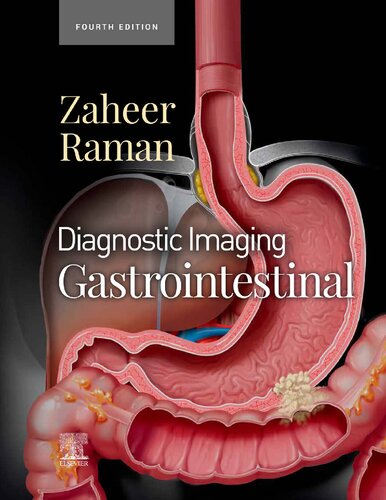
Diagnostic Imaging Gastrointestinal 4th Edition by Atif Zaheer, Siva P Raman ISBN 9780323824989 0323824986
$50.00 Original price was: $50.00.$25.00Current price is: $25.00.
Diagnostic Imaging Gastrointestinal 4th Edition by Atif Zaheer, Siva P Raman – Ebook PDF Instant Download/Delivery: 9780323824989, 0323824986
Full download Diagnostic Imaging Gastrointestinal 4th Edition after payment

Product details:
ISBN 10: 0323824986
ISBN 13: 9780323824989
Author: Atif Zaheer, Siva P Raman
Covering the entire spectrum of this fast-changing field, Diagnostic Imaging: Gastrointestinal, fourth edition, is an invaluable resource for gastrointestinal radiologists, general radiologists, and trainees—anyone who requires an easily accessible, highly visual reference on today’s GI imaging. Drs. Siva P. Raman, Atif Zaheer, and their team of highly regarded experts provide up-to-date information on recent advances in technology and the understanding of GI diseases and disorders to help you make informed decisions at the point of care. The text is lavishly illustrated, delineated, and referenced, making it a useful learning tool as well as a handy reference for daily practice.
-
Serves as a one-stop resource for key concepts and information on gastrointestinal imaging, including a wealth of new material and content updates throughout
-
Features more than 2,900 illustrations (multiplanar CT, sonography, MR, and PET/CT; clinical photos; radiologic images; histologic images; H&E stains; and full-color illustrations) as well as an additional 3K digital-only images and new video clips
-
Features updates from cover to cover including new information on MRI imaging of rectal cancer, iron quantification, and MRI protocols; new cases and images, and new staging details and diagrams
-
Contains new chapters on treatment response criteria for systemic conditions (RECIST, irRECIST, etc.), dual energy CT for pancreas, vascular abnormalities, MR elastography of the liver, pretransplant liver evaluation, and more
-
Covers all aspects of GI imaging, including pathophysiology, imaging findings, and disease management options such as the radiologist’s role in evaluating patients for bariatric surgery, antireflux procedures, esophageal and bowel resections, and more
-
Uses bulleted, succinct text and highly templated chapters for quick comprehension of essential information at the point of care
Diagnostic Imaging Gastrointestinal 4th Edition Table of contents:
SECTION 1: ABDOMINAL MANIFESTATIONS OF SYSTEMIC CONDITIONS
INTRODUCTION AND OVERVIEW
Chapter 1: Imaging Approach to Abdominal Manifestations of Systemic Conditions
Main Text
Image Gallery
INFECTION
Chapter 2: HIV/AIDS
Key Facts
Key Images
Main Text
Image Gallery
Chapter 3: Tuberculosis
Key Facts
Key Images
Main Text
Image Gallery
Chapter 4: Mononucleosis
Key Facts
Key Images
Main Text
Image Gallery
METABOLIC OR INHERITED
Chapter 5: Cystic Fibrosis
Key Facts
Key Images
Main Text
Image Gallery
Chapter 6: Sickle Cell Anemia
Key Facts
Key Images
Main Text
Image Gallery
Chapter 7: Amyloidosis
Key Facts
Key Images
Main Text
Image Gallery
Chapter 8: Sarcoidosis
Key Facts
Key Images
Main Text
Image Gallery
VASCULAR DISORDERS
Chapter 9: Systemic Hypotension
Key Facts
Key Images
Main Text
Image Gallery
Chapter 10: Superior Vena Cava Obstruction
Key Facts
Key Images
Main Text
Image Gallery
Chapter 11: Vasculitis
Key Facts
Key Images
Main Text
Image Gallery
TRAUMA
Chapter 12: Foreign Bodies
Key Facts
Key Images
Main Text
Image Gallery
Chapter 13: Barotrauma
Key Facts
Key Images
Main Text
TRANSPLANTATION
Chapter 14: Posttransplant Lymphoproliferative Disorder
Key Facts
Key Images
Main Text
Image Gallery
MALIGNANT NEOPLASMS
Chapter 15: Leukemia and Lymphoma
Key Facts
Key Images
Main Text
Image Gallery
Chapter 16: Metastatic Melanoma
Key Facts
Key Images
Main Text
Image Gallery
Chapter 17: Kaposi Sarcoma
Key Facts
Key Images
Main Text
Image Gallery
Chapter 18: Treatment Response Assessment
Key Facts
Key Images
Main Text
Tables
Image Gallery
SECTION 2: PERITONEUM, MESENTERY, AND ABDOMINAL WALL
INTRODUCTION AND OVERVIEW
Chapter 19: Imaging Approach to Peritoneum, Mesentery, and Abdominal Wall
Main Text
Image Gallery
INFECTION
Chapter 20: Abdominal Abscess
Key Facts
Key Images
Main Text
Image Gallery
INFLAMMATION
Chapter 21: Peritonitis
Key Facts
Key Images
Main Text
Image Gallery
Chapter 22: Sclerosing Mesenteritis
Key Facts
Key Images
Main Text
Image Gallery
DEGENERATIVE
Chapter 23: Ascites
Key Facts
Key Images
Main Text
Image Gallery
Chapter 24: Omental Infarct
Key Facts
Key Images
Main Text
Image Gallery
EXTERNAL HERNIAS
Chapter 25: Inguinal Hernia
Key Facts
Key Images
Main Text
Image Gallery
Chapter 26: Femoral Hernia
Key Facts
Key Images
Main Text
Image Gallery
Chapter 27: Obturator Hernia
Key Facts
Key Images
Main Text
Image Gallery
Chapter 28: Ventral Hernia
Key Facts
Key Images
Main Text
Image Gallery
Chapter 29: Spigelian Hernia
Key Facts
Key Images
Main Text
Image Gallery
Chapter 30: Lumbar Hernia
Key Facts
Key Images
Main Text
Image Gallery
Chapter 31: Umbilical Hernia
Key Facts
Key Images
Main Text
Image Gallery
INTERNAL HERNIAS
Chapter 32: Paraduodenal Hernia
Key Facts
Key Images
Main Text
Image Gallery
Chapter 33: Transmesenteric Postoperative Hernia
Key Facts
Key Images
Main Text
Image Gallery
Chapter 34: Bochdalek Hernia
Key Facts
Key Images
Main Text
Image Gallery
Chapter 35: Morgagni Hernia
Key Facts
Key Images
Main Text
Image Gallery
VASCULAR DISORDERS
Chapter 36: Portal Hypertension and Varices
Key Facts
Key Images
Main Text
Image Gallery
Chapter 37: Visceral Aneurysms and Pseudoaneurysms
Key Facts
Key Images
Main Text
Image Gallery
TRAUMA
Chapter 38: Traumatic Abdominal Wall Hernia
Key Facts
Key Images
Main Text
Image Gallery
Chapter 39: Traumatic Diaphragmatic Rupture
Key Facts
Key Images
Main Text
Image Gallery
TREATMENT RELATED
Chapter 40: Postoperative State, Abdomen
Key Facts
Key Images
Main Text
Image Gallery
Chapter 41: Abdominal Incision and Injection Sites
Key Facts
Key Images
Main Text
Image Gallery
Chapter 42: Peritoneal Inclusion Cyst
Key Facts
Key Images
Main Text
Image Gallery
BENIGN NEOPLASMS
Chapter 43: Lymphangioma (Mesenteric Cyst)
Key Facts
Key Images
Main Text
Image Gallery
Chapter 44: Desmoid
Key Facts
Key Images
Main Text
Image Gallery
MALIGNANT NEOPLASMS
Chapter 45: Abdominal Mesothelioma
Key Facts
Key Images
Main Text
Image Gallery
Chapter 46: Peritoneal Metastases
Key Facts
Key Images
Main Text
Image Gallery
Chapter 47: Pseudomyxoma Peritonei
Key Facts
Key Images
Main Text
Image Gallery
MISCELLANEOUS
Chapter 48: Eventration and Paralysis of Diaphragm
Key Facts
Key Images
Main Text
Image Gallery
Chapter 49: Vicarious Excretion
Key Facts
Key Images
Main Text
Image Gallery
SECTION 3: ESOPHAGUS
INTRODUCTION AND OVERVIEW
Chapter 50: Imaging Approach to Esophagus
Main Text
Image Gallery
INFECTION
Chapter 51: Candida Esophagitis
Key Facts
Key Images
Main Text
Image Gallery
Chapter 52: Viral Esophagitis
Key Facts
Key Images
Main Text
Chapter 53: Chagas Disease
Key Facts
Key Images
Main Text
Image Gallery
INFLAMMATION
Chapter 54: Reflux Esophagitis
Key Facts
Key Images
Main Text
Image Gallery
Chapter 55: Barrett Esophagus
Key Facts
Key Images
Main Text
Image Gallery
Chapter 56: Caustic Esophagitis
Key Facts
Key Images
Main Text
Image Gallery
Chapter 57: Drug-Induced Esophagitis
Key Facts
Key Images
Main Text
Image Gallery
Chapter 58: Radiation Esophagitis
Key Facts
Key Images
Main Text
Image Gallery
Chapter 59: Eosinophilic Esophagitis
Key Facts
Key Images
Main Text
Chapter 60: Epidermolysis and Pemphigoid
Key Facts
Key Images
Main Text
DEGENERATIVE
Chapter 61: Esophageal Webs
Key Facts
Key Images
Main Text
Image Gallery
Chapter 62: Cricopharyngeal Achalasia
Key Facts
Key Images
Main Text
Image Gallery
Chapter 63: Esophageal Achalasia
Key Facts
Key Images
Main Text
Image Gallery
Chapter 64: Esophageal Motility Disturbances
Key Facts
Key Images
Main Text
Image Gallery
Chapter 65: Esophageal Scleroderma
Key Facts
Key Images
Main Text
Image Gallery
Chapter 66: Schatzki Ring
Key Facts
Key Images
Main Text
Image Gallery
Chapter 67: Hiatal Hernia
Key Facts
Key Images
Main Text
Image Gallery
VASCULAR DISORDERS
Chapter 68: Esophageal Varices
Key Facts
Key Images
Main Text
Image Gallery
ESOPHAGEAL DIVERTICULA
Chapter 69: Zenker Diverticulum
Key Facts
Key Images
Main Text
Image Gallery
Chapter 70: Intramural Pseudodiverticulosis
Key Facts
Key Images
Main Text
Image Gallery
Chapter 71: Traction Diverticulum
Key Facts
Key Images
Main Text
Image Gallery
Chapter 72: Pulsion Diverticulum
Key Facts
Key Images
Main Text
Image Gallery
TRAUMA
Chapter 73: Esophageal Foreign Body
Key Facts
Key Images
Main Text
Image Gallery
Chapter 74: Esophageal Perforation
Key Facts
Key Images
Main Text
Image Gallery
Chapter 75: Boerhaave Syndrome
Key Facts
Key Images
Main Text
Image Gallery
TREATMENT RELATED
Chapter 76: Esophagectomy: Ivor Lewis and Other Procedures
Key Facts
Key Images
Main Text
Image Gallery
BENIGN NEOPLASMS
Chapter 77: Intramural Benign Esophageal Tumors
Key Facts
Key Images
Main Text
Image Gallery
Chapter 78: Fibrovascular Polyp
Key Facts
Key Images
Main Text
Image Gallery
Chapter 79: Esophageal Inflammatory Polyp
Key Facts
Key Images
Main Text
MALIGNANT NEOPLASMS
Chapter 80: Esophageal Carcinoma
Key Facts
Key Images
Main Text
Image Gallery
Chapter 81: Esophageal Metastases and Lymphoma
Key Facts
Key Images
Main Text
Image Gallery
SECTION 4: STOMACH
INTRODUCTION AND OVERVIEW
Chapter 82: Imaging Approach to Stomach
Main Text
Image Gallery
CONGENITAL
Chapter 83: Gastric Diverticulum
Key Facts
Key Images
Main Text
Image Gallery
INFLAMMATION
Chapter 84: Gastritis
Key Facts
Key Images
Main Text
Image Gallery
Chapter 85: Gastric Ulcer
Key Facts
Key Images
Main Text
Image Gallery
Chapter 86: Zollinger-Ellison Syndrome
Key Facts
Key Images
Main Text
Image Gallery
Chapter 87: Ménétrier Disease
Key Facts
Key Images
Main Text
Image Gallery
Chapter 88: Caustic Gastroduodenal Injury
Key Facts
Key Images
Main Text
Image Gallery
DEGENERATIVE
Chapter 89: Gastroparesis
Key Facts
Key Images
Main Text
Chapter 90: Gastric Bezoar
Key Facts
Key Images
Main Text
Image Gallery
Chapter 91: Gastric Volvulus
Key Facts
Key Images
Main Text
Image Gallery
TREATMENT RELATED
Chapter 92: Iatrogenic Injury: Feeding Tubes
Key Facts
Key Images
Main Text
Image Gallery
People also search for Diagnostic Imaging Gastrointestinal 4th Edition:
diagnostic imaging gastrointestinal 4th edition
diagnostic imaging gastrointestinal e book
diagnostic imaging pathways gastrointestinal bleeding obscure
diagnostic radiology gastrointestinal and hepatobiliary imaging
diagnostic imaging examples
Tags: Atif Zaheer, Siva P Raman, Diagnostic Imaging Gastrointestinal
You may also like…
Medicine - Pediatrics
Diagnostic Imaging: Pediatric Neuroradiology 3rd Edition Kevin R. Moore
Medicine - Others
Medicine - Others
Diagnostic Imaging: Obstetrics 4th Edition Paula J. Woodward Md
Medicine - Others
Medicine - Oncology
Diagnostic Imaging Oncology by Akram M. Shaaban 9780323661133 0323661130
Medicine - Radiology
Diagnostic Imaging Musculoskeletal Trauma Donna G. Blankenbaker


